Patient undergoing echocardiography
Numéro d’image : 12068607
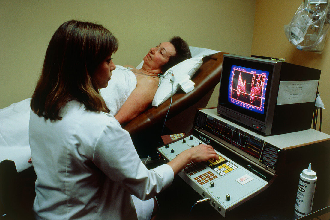
| View of a patient undergoing an echocardiography examination. This method produces an image of the heart by using ultrasound,which are inaudible and harmless high-frequency sound waves. It is mostly used to detect functional abnormalities of the heart wall,the heart valves and the large blood vessels connected to the heart. The sound waves are transmitted by a set of transducers placed on the chest of the patient. The echoes from the different transducers are detected,amplified and then combined to produce a two-dimensional image on a TV screen | |
| Licence : | Droits gérés |
| Crédit: | Science Photo Library / Leavines, Susan |
| Taille de l’image : | 5287 px × 3536 px |
| Model Release : | Le droit n'est pas encore disponible. Merci de nous contacter avant utilisation. |
| Property Release : | Non requis |
| Restrictions : |
|
Prix pour cette image À partir de 45 €
Produit vendu
(Calendrier, Carte postale, Carte de vœux, Impression sur textile, Packaging etc)
À partir de 45 €
Usage commercial
(Affichage, Annonce presse, Annonce TV, Carte, Digital - hors rés. sociaux, Digital - rés. sociaux etc)
À partir de 45 €
Éditorial
(Digital, Journal, Livre, Livre pratique, Magazine, Télévision etc)
À partir de 60 €
Usage non-commercial
(Digital - hors rés. sociaux, Digital - rés. sociaux etc)
À partir de 120 €