Knee with fracture of distal femur
Numéro d’image : 12068256
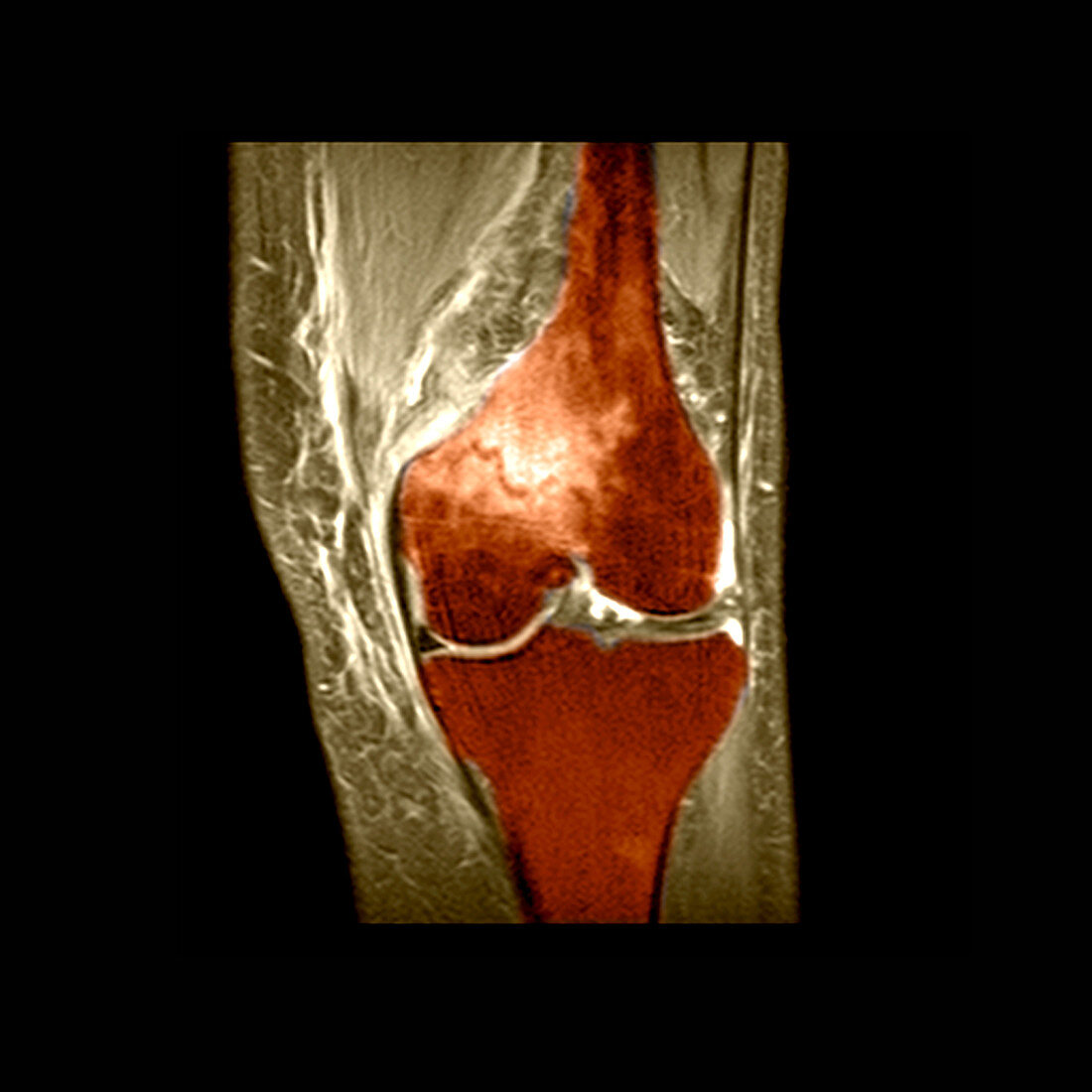
| colour enhanced MRI (magnetic resonance imaging) scan,showing the frontal view of knee with a fracture of the distal femur. The fracture is the jagged dark line surrounded by bright signal in the distal femur. The bright signal represents bone marrow edema secondary to an acute fracture. The bones comprising the knee (femur and tibia) are cushioned by the menisci (articular fibrocartilage). These are seen as triangular black densities on each side of the joint and between the bones. The signal which gives signal to the inside of the bone is from the fatty bone marrow | |
| Licence : | Droits gérés |
| Crédit: | Science Photo Library / Living Art Enterprises, LLC |
| Taille de l’image : | 3600 px × 3600 px |
| Model Release : | Non requis |
| Property Release : | Non requis |
| Restrictions : |
|
Prix pour cette image À partir de 45 €
Produit vendu
(Calendrier, Carte postale, Carte de vœux, Impression sur textile, Packaging etc)
À partir de 45 €
Usage commercial
(Affichage, Annonce presse, Annonce TV, Carte, Digital - hors rés. sociaux, Digital - rés. sociaux etc)
À partir de 45 €
Éditorial
(Digital, Journal, Livre, Livre pratique, Magazine, Télévision etc)
À partir de 60 €
Usage non-commercial
(Digital - hors rés. sociaux, Digital - rés. sociaux etc)
À partir de 120 €
Mots clés
- articulation,
- articulations,
- blessure,
- blessure au genou,
- désordre,
- diagnostic,
- diagnostique,
- état,
- fémur distal,
- fémurs,
- fracture,
- fractures,
- genou,
- genoux,
- I.R.M.,
- imagerie médicale,
- imagerie par résonance magnétique,
- imagerie par résonnance magnétique,
- IRM,
- maladie,
- médecine,
- médical,
- médicale,
- os,
- soins de santé,
- système squelettique,
- trouble