Computer Reconstruction of the Knee
Numéro d’image : 12068251
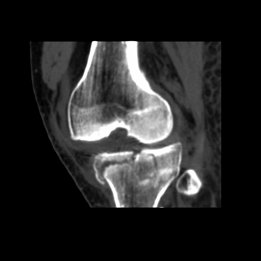
| This image is a computer generated reconstruction of the knee demonstrating a comminuted (multiple fragments) fracture extending through the tibial plateau which is one of the bones of the knee. The other major bone comprising the knee joint is the distal femur. A smaller bone the patella is the 3rd bone making up the knee. Computed Tomography (CT) or computed axial tomography (CAT) scans generate special types of images which utilize x-rays for their creation. CT scans obtain cross sectional images of different body parts. Complex fractures or fractures which are not adequately imaged by plain X-rays are better evaluated with CT scans. It is often helpful to determine if a fracture extends to a joint,as this information may alter the way a person is treated. This case illustrates a very severe fracture which does involve the knee joint | |
| Licence : | Droits gérés |
| Crédit: | Science Photo Library / Living Art Enterprises, LLC |
| Taille de l’image : | 4800 px × 4800 px |
| Model Release : | Non requis |
| Property Release : | Non requis |
| Restrictions : |
|
Prix pour cette image À partir de 45 €
Produit vendu
(Calendrier, Carte postale, Carte de vœux, Impression sur textile, Packaging etc)
À partir de 45 €
Usage commercial
(Affichage, Annonce presse, Annonce TV, Carte, Digital - hors rés. sociaux, Digital - rés. sociaux etc)
À partir de 45 €
Éditorial
(Digital, Journal, Livre, Livre pratique, Magazine, Télévision etc)
À partir de 60 €
Usage non-commercial
(Digital - hors rés. sociaux, Digital - rés. sociaux etc)
À partir de 120 €
Mots clés
- articulation,
- articulations,
- balayage,
- balayages,
- blessure,
- fémur,
- fémur distal,
- fémurs,
- généré par ordinateur,
- genou,
- genoux,
- imagerie médicale,
- images médicales,
- médecine,
- médical,
- médicale,
- numérisation,
- os,
- os de jambe,
- os de la jambe,
- os du genou,
- patella,
- reconstruction,
- reconstructions,
- rotule,
- scanner,
- scanner CT,
- scanographie,
- scans,
- soins de santé,
- TDM,
- tibia,
- tomodensitométrie,
- tomographie assistée par ordinateur