MRI of Otomastoiditis and Sphenoid Sinusitis
Numéro d’image : 12068161
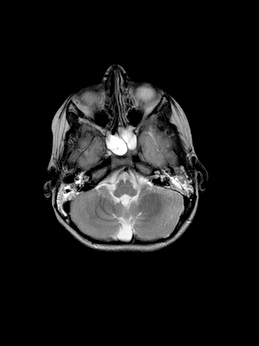
| This T2 weighted axial (cross sectional) MRI image of the head beautifully demonstrates bilateral otomastoiditis and sphenoid sinusitis. There is extensive increased signal intensity (looks white) in the mastoid air cells and middle ear clefts,bilaterally. There is also complete opacification of the sphenoid sinus,which also demonstrates prominent abnormal increased signal intensity (looks white) | |
| Licence : | Droits gérés |
| Crédit: | Science Photo Library / Living Art Enterprises, LLC |
| Taille de l’image : | 2700 px × 3600 px |
| Model Release : | Non requis |
| Property Release : | Non requis |
| Restrictions : |
|
Prix pour cette image À partir de 45 €
Produit vendu
(Calendrier, Carte postale, Carte de vœux, Impression sur textile, Packaging etc)
À partir de 45 €
Usage commercial
(Affichage, Annonce presse, Annonce TV, Carte, Digital - hors rés. sociaux, Digital - rés. sociaux etc)
À partir de 45 €
Éditorial
(Digital, Journal, Livre, Livre pratique, Magazine, Télévision etc)
À partir de 60 €
Usage non-commercial
(Digital - hors rés. sociaux, Digital - rés. sociaux etc)
À partir de 120 €