MRI of Sturge-Weber Syndrome
Numéro d’image : 12068157
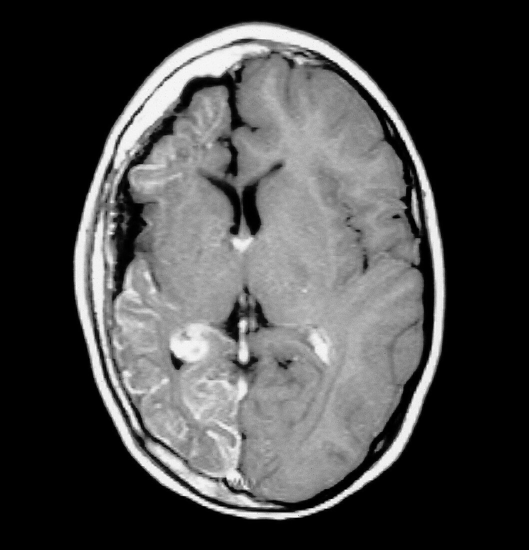
| This axial (cross sectional) contrast enhanced MRI image of the brain shows findings consistent with Sturge-Weber syndrome,also known as encephalo-trigeminal angiomatosis. This is characterized by a pial leptomeningeal venous angiomatosis,cerebral hemiatrophy on the side of the venous abnormality,seizures and a port-wine stain (nevus) in the distribution of the trigeminal nerve ipsilateral to the intracranial abnormality | |
| Licence : | Droits gérés |
| Crédit: | Science Photo Library / Living Art Enterprises, LLC |
| Taille de l’image : | 3895 px × 4045 px |
| Model Release : | Non requis |
| Property Release : | Non requis |
| Restrictions : |
|
Prix pour cette image À partir de 45 €
Produit vendu
(Calendrier, Carte postale, Carte de vœux, Impression sur textile, Packaging etc)
À partir de 45 €
Usage commercial
(Affichage, Annonce presse, Annonce TV, Carte, Digital - hors rés. sociaux, Digital - rés. sociaux etc)
À partir de 45 €
Éditorial
(Digital, Journal, Livre, Livre pratique, Magazine, Télévision etc)
À partir de 60 €
Usage non-commercial
(Digital - hors rés. sociaux, Digital - rés. sociaux etc)
À partir de 120 €