PET scans normal and schizophrenic brains
Numéro d’image : 12068118
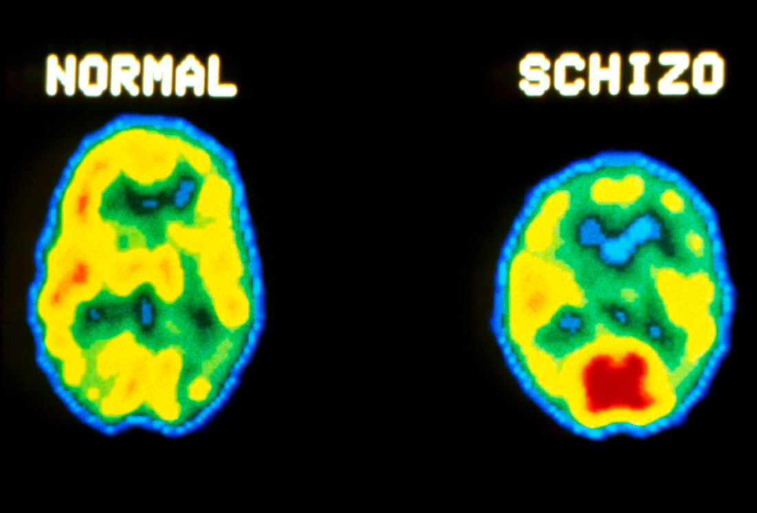
| Two axial PET scans of the brain,comparing a normal subject (left) with a person suffering from schizophrenia. PET scans (Positron Emission Tomography) obtain details of the function of tissues (as opposed to structural aspects resolved by NMR and CAT scans) using tracers labelled with a short-lived radioisotope. Here,a glucose analogue labelled with oxygen-11 was injected into the bloodstream immediately prior to taking the scans,crossing the blood-brain barrier to participate in glucose metabolism in the brain. Photons emitted as the positrons decay are detected & processed to give colour-coded images | |
| Licence : | Droits gérés |
| Crédit: | Science Photo Library / US National Institute of Health |
| Taille de l’image : | 3713 px × 2516 px |
| Model Release : | Non requis |
| Property Release : | Non requis |
| Restrictions : |
|
Prix pour cette image À partir de 45 €
Produit vendu
(Calendrier, Carte postale, Carte de vœux, Impression sur textile, Packaging etc)
À partir de 45 €
Usage commercial
(Affichage, Annonce presse, Annonce TV, Carte, Digital - hors rés. sociaux, Digital - rés. sociaux etc)
À partir de 45 €
Éditorial
(Digital, Journal, Livre, Livre pratique, Magazine, Télévision etc)
À partir de 60 €
Usage non-commercial
(Digital - hors rés. sociaux, Digital - rés. sociaux etc)
À partir de 120 €