CT of Hiatal Hernia and Liver Cyst
Numéro d’image : 12067772
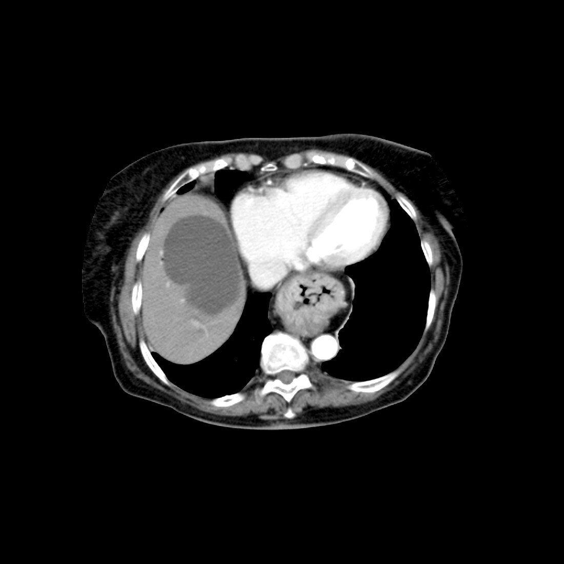
| This axial (cross sectional) CT through the lower chest,with intravenous contrast,shows a large hiatal hernia. You can visualise the rugal folds within the hiatal hernia,indicating that this is in fact the stomach. This person suffered from gastro-esophageal reflux disease (GERD). Also note the large,benign cyst within the liver | |
| Licence : | Droits gérés |
| Crédit: | Science Photo Library / Living Art Enterprises, LLC |
| Taille de l’image : | 3600 px × 3600 px |
| Model Release : | Non requis |
| Property Release : | Non requis |
| Restrictions : |
|
Prix pour cette image À partir de 45 €
Produit vendu
(Calendrier, Carte postale, Carte de vœux, Impression sur textile, Packaging etc)
À partir de 45 €
Usage commercial
(Affichage, Annonce presse, Annonce TV, Carte, Digital - hors rés. sociaux, Digital - rés. sociaux etc)
À partir de 45 €
Éditorial
(Digital, Journal, Livre, Livre pratique, Magazine, Télévision etc)
À partir de 60 €
Usage non-commercial
(Digital - hors rés. sociaux, Digital - rés. sociaux etc)
À partir de 120 €