Fibrous dysplasia
Numéro d’image : 12067711
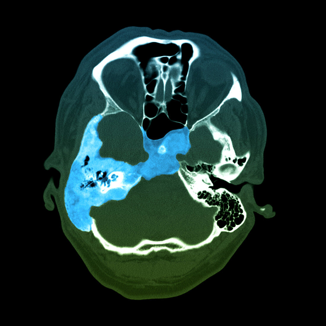
| This colour enhanced axial (cross section) CT image of the head shows a typical appearance of fibrous dysplasia (blue),described as a ground glass enlargement of the involved bony structures. In this case there is involvement of the petrous temporal bone housing the tiny bones of the middle ear (ossicles). There is also involvement of the mastoid region on the right and the basisphenoid bone | |
| Licence : | Droits gérés |
| Crédit: | Science Photo Library / Living Art Enterprises, LLC |
| Taille de l’image : | 3600 px × 3600 px |
| Model Release : | Non requis |
| Property Release : | Non requis |
| Restrictions : |
|
Prix pour cette image À partir de 45 €
Produit vendu
(Calendrier, Carte postale, Carte de vœux, Impression sur textile, Packaging etc)
À partir de 45 €
Usage commercial
(Affichage, Annonce presse, Annonce TV, Carte, Digital - hors rés. sociaux, Digital - rés. sociaux etc)
À partir de 45 €
Éditorial
(Digital, Journal, Livre, Livre pratique, Magazine, Télévision etc)
À partir de 60 €
Usage non-commercial
(Digital - hors rés. sociaux, Digital - rés. sociaux etc)
À partir de 120 €
Mots clés
- amélioration de couleur,
- cerveau,
- couleur,
- couleur améliorée,
- couleur augmentée,
- désordre,
- élargissement,
- état,
- image médicale,
- imagerie médicale,
- maladie,
- médecine,
- médical,
- médicale,
- neuro-imagerie,
- neuroimagerie,
- oreille moyenne,
- os,
- osselet,
- scanner,
- science,
- soins de santé,
- tomodensitogramme,
- tomodensitométrie,
- trouble