Severe bullous emphysema
Numéro d’image : 12067676
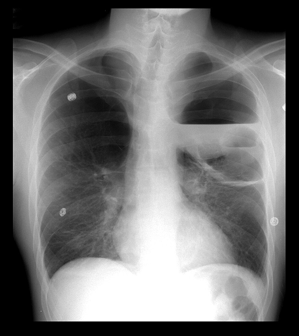
| This frontal chest x-ray shows an example of severe bullous emphysema in the left upper lobe (to your right) and,to a lesser degree,the right upper lobe (to your left). In the left upper lobe there are multiple air-fluid levels reflecting infected bullae | |
| Licence : | Droits gérés |
| Crédit: | Science Photo Library / Living Art Enterprises, LLC |
| Taille de l’image : | 3300 px × 3713 px |
| Model Release : | Non requis |
| Property Release : | Non requis |
| Restrictions : |
|
Prix pour cette image À partir de 45 €
Produit vendu
(Calendrier, Carte postale, Carte de vœux, Impression sur textile, Packaging etc)
À partir de 45 €
Usage commercial
(Affichage, Annonce presse, Annonce TV, Carte, Digital - hors rés. sociaux, Digital - rés. sociaux etc)
À partir de 45 €
Éditorial
(Digital, Journal, Livre, Livre pratique, Magazine, Télévision etc)
À partir de 60 €
Usage non-commercial
(Digital - hors rés. sociaux, Digital - rés. sociaux etc)
À partir de 120 €
Mots clés
- asthma,
- asthme,
- bronchite,
- bronchitis,
- désordre,
- emphysema,
- emphysème,
- emphysème bulleux,
- état,
- image médicale,
- imagerie médicale,
- infection du poumon,
- maladie,
- maladie pulmonaire,
- médecine,
- médical,
- médicale,
- MPOC,
- radiographie,
- radiographie de la poitrine,
- radiographie du buste,
- radiographie du thorax,
- radiographie thoracique,
- science,
- soins de santé,
- trouble