Depression: 2 axial PET scans of the brain
Numéro d’image : 12067629
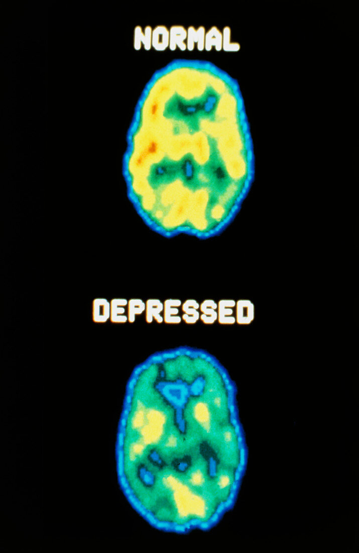
| Depression. Two axial PET scans of the brain,comparing a normal subject (top) with a person suffering from a depressive illness. The depressed patient shows lowered brain activity. PET scans (Positron Emission Tomography) obtain details of the function of tissues using tracers labelled with a short-lived radioisotope. Here,a glucose analogue labelled with oxygen-11 is injected into the bloodstream immediately prior to taking the scan,crossing the blood-brain barrier to participate in glucose metabolism in the brain. Positrons emitted are detected and data processed to provide the colour-coded images | |
| Licence : | Droits gérés |
| Crédit: | Science Photo Library / US National Institute of Health |
| Taille de l’image : | 3206 px × 4935 px |
| Model Release : | Non requis |
| Property Release : | Non requis |
| Restrictions : |
|
Prix pour cette image À partir de 45 €
Produit vendu
(Calendrier, Carte postale, Carte de vœux, Impression sur textile, Packaging etc)
À partir de 45 €
Usage commercial
(Affichage, Annonce presse, Annonce TV, Carte, Digital - hors rés. sociaux, Digital - rés. sociaux etc)
À partir de 45 €
Éditorial
(Digital, Journal, Livre, Livre pratique, Magazine, Télévision etc)
À partir de 60 €
Usage non-commercial
(Digital - hors rés. sociaux, Digital - rés. sociaux etc)
À partir de 120 €