Stroke
Numéro d’image : 12067579
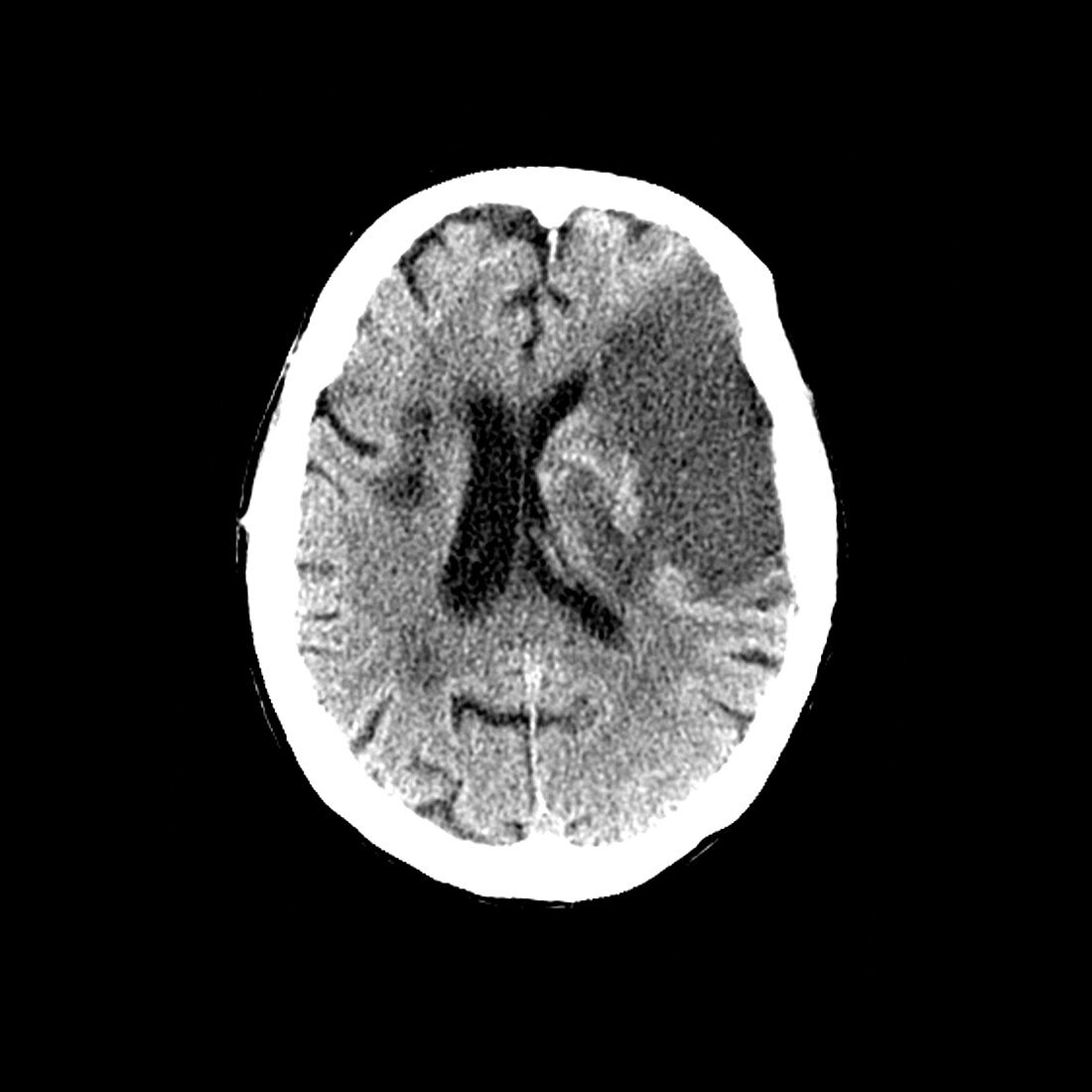
| Computerized axial tomography image of the brain of a patient with both an inability to speak and right-side weakness. The large wedge-shaped area of lower density on the right is a large acute stroke. The smaller area of low density on the left is an old stroke. The central black cavities to surrounding the midline are the lateral ventricles filled with cerebral spinal fluid (CSF) | |
| Licence : | Droits gérés |
| Crédit: | Science Photo Library / Living Art Enterprises, LLC |
| Taille de l’image : | 3600 px × 3600 px |
| Model Release : | Non requis |
| Property Release : | Non requis |
| Restrictions : |
|
Prix pour cette image À partir de 45 €
Produit vendu
(Calendrier, Carte postale, Carte de vœux, Impression sur textile, Packaging etc)
À partir de 45 €
Usage commercial
(Affichage, Annonce presse, Annonce TV, Carte, Digital - hors rés. sociaux, Digital - rés. sociaux etc)
À partir de 45 €
Éditorial
(Digital, Journal, Livre, Livre pratique, Magazine, Télévision etc)
À partir de 60 €
Usage non-commercial
(Digital - hors rés. sociaux, Digital - rés. sociaux etc)
À partir de 120 €
Mots clés
- A.V.C.,
- accident vasculaire cérébral,
- AVC,
- balayages,
- CAT-scan,
- cerveau,
- chat,
- coup important,
- désordre,
- état,
- fluide cérébrospinal,
- liquide céphalo-rachidien,
- liquide céphalorachidien,
- maladie,
- médecine,
- médical,
- médicale,
- neuro-imagerie,
- neuroimagerie,
- scanner,
- scans,
- soins de santé,
- T.D.M.,
- TDM,
- tomodensitogramme,
- tomodensitométrie,
- tomodensitométrie axiale,
- tomographe,
- tomographie,
- trouble,
- vaisseau sanguin,
- vasculaire