Angiogram of cerebral arteriovenous malformation
Numéro d’image : 12067502
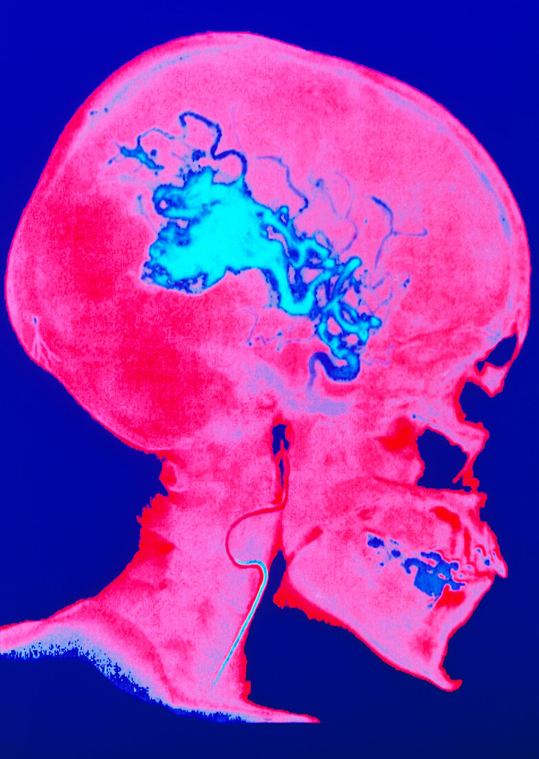
| Brain arteriovenous malformation. False-colour angiogram of the human head in lateral view,showing an arteriovenous malformation (AVM) in the brain. This is outlined by a radio-opaque contrast medium (light blue) and seen on X-ray. Here,the common carotid artery at the base of the neck (blue line) has a catheter & internal guide- wire advancing within it to the site of the AVM. It will deliver an embolic foam material to occlude the blood vessel malformations. This condition is also known as angioma: a knot of dis- tended blood vessels overlying and compressing the brain surface. It commonly causes epilepsy. A sub-arachnoid haemorrhage may also result | |
| Licence : | Droits gérés |
| Crédit: | Science Photo Library / Lunagrafix |
| Taille de l’image : | 3543 px × 4984 px |
| Model Release : | Non requis |
| Property Release : | Non requis |
| Restrictions : |
|
Prix pour cette image À partir de 45 €
Produit vendu
(Calendrier, Carte postale, Carte de vœux, Impression sur textile, Packaging etc)
À partir de 45 €
Usage commercial
(Affichage, Annonce presse, Annonce TV, Carte, Digital - hors rés. sociaux, Digital - rés. sociaux etc)
À partir de 45 €
Éditorial
(Digital, Journal, Livre, Livre pratique, Magazine, Télévision etc)
À partir de 60 €
Usage non-commercial
(Digital - hors rés. sociaux, Digital - rés. sociaux etc)
À partir de 120 €