LM of metastatic carcinoma (cancer) of the liver
Numéro d’image : 12067335
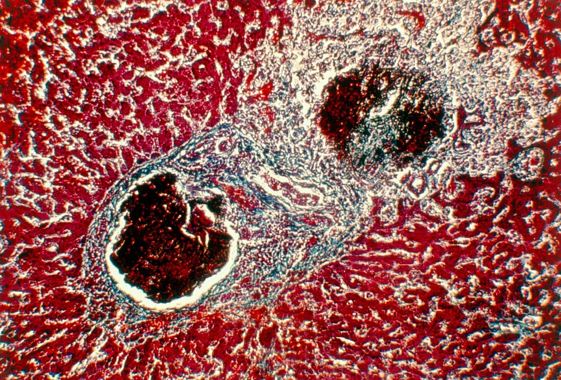
| Metastatic liver carcinoma. Light micrograph of a section through the human liver,showing secondary (metastasised) carcinoma. Two cancerous tumours (darkly stained) are seen. They consist of loosely arranged cancer cells undergoing mitotic activity (cell division). Cell growth appears random and chaotic. Healthy liver tissue (red) can be seen around these tumours. Secondary liver carcinoma spreads from other primary sites in the body,notably the stomach,pancreas,intestine and skin. Being a blood filtering organ,the liver is a common site of secondary (metastatic) cancers Magnification: x40 at 35mm size | |
| Licence : | Droits gérés |
| Crédit: | Science Photo Library / Edward, Kenneth / Biografx |
| Taille de l’image : | 5138 px × 3483 px |
| Model Release : | Non requis |
| Property Release : | Non requis |
| Restrictions : |
|
Prix pour cette image À partir de 45 €
Produit vendu
(Calendrier, Carte postale, Carte de vœux, Impression sur textile, Packaging etc)
À partir de 45 €
Usage commercial
(Affichage, Annonce presse, Annonce TV, Carte, Digital - hors rés. sociaux, Digital - rés. sociaux etc)
À partir de 45 €
Éditorial
(Digital, Journal, Livre, Livre pratique, Magazine, Télévision etc)
À partir de 60 €
Usage non-commercial
(Digital - hors rés. sociaux, Digital - rés. sociaux etc)
À partir de 120 €