Temporal Lobe Cavernous Malformations
Numéro d’image : 12067245
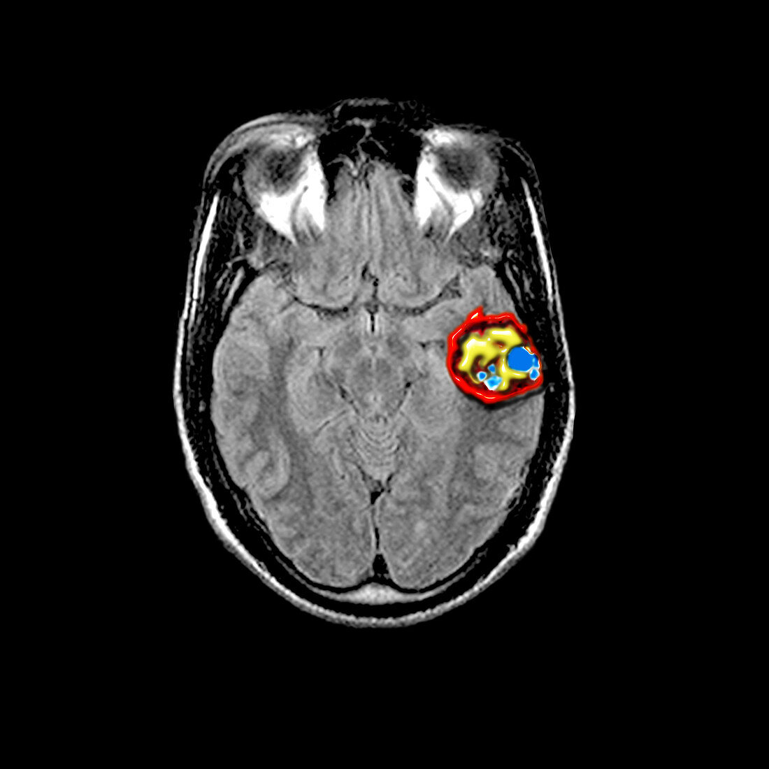
| Coloured cross-sectional MRI image of the head through the temporal lobes,midbrain,and inferior frontal lobes. There is a large cavernous malformation in the left temporal lobe which is on the right side of the image. Cavernous malformations are vascular lesions which are composed of clusters of abnormally dilated capillaries. These lesions bleed and are composted of blood of varying ages. The red ring around the lesion represents chronic (old) blood. Inside of the lesion blood of varying ages is represented by irregular shaped areas of different colors. These abnormal lesions can cause symptoms as a result of their location near or in important brain tissue. They can also expand (enlarge) by oozing blood around their edges. When these lesions cause significant symptoms they can be removed by a neurosurgeon if they are located in a surgically accesible region of the brain | |
| Licence : | Droits gérés |
| Crédit: | Science Photo Library / Living Art Enterprises, LLC |
| Taille de l’image : | 4200 px × 4200 px |
| Model Release : | Non requis |
| Property Release : | Non requis |
| Restrictions : |
|
Prix pour cette image À partir de 45 €
Produit vendu
(Calendrier, Carte postale, Carte de vœux, Impression sur textile, Packaging etc)
À partir de 45 €
Usage commercial
(Affichage, Annonce presse, Annonce TV, Carte, Digital - hors rés. sociaux, Digital - rés. sociaux etc)
À partir de 45 €
Éditorial
(Digital, Journal, Livre, Livre pratique, Magazine, Télévision etc)
À partir de 60 €
Usage non-commercial
(Digital - hors rés. sociaux, Digital - rés. sociaux etc)
À partir de 120 €