Benign Pilocytic Astrocytoma
Numéro d’image : 12067114
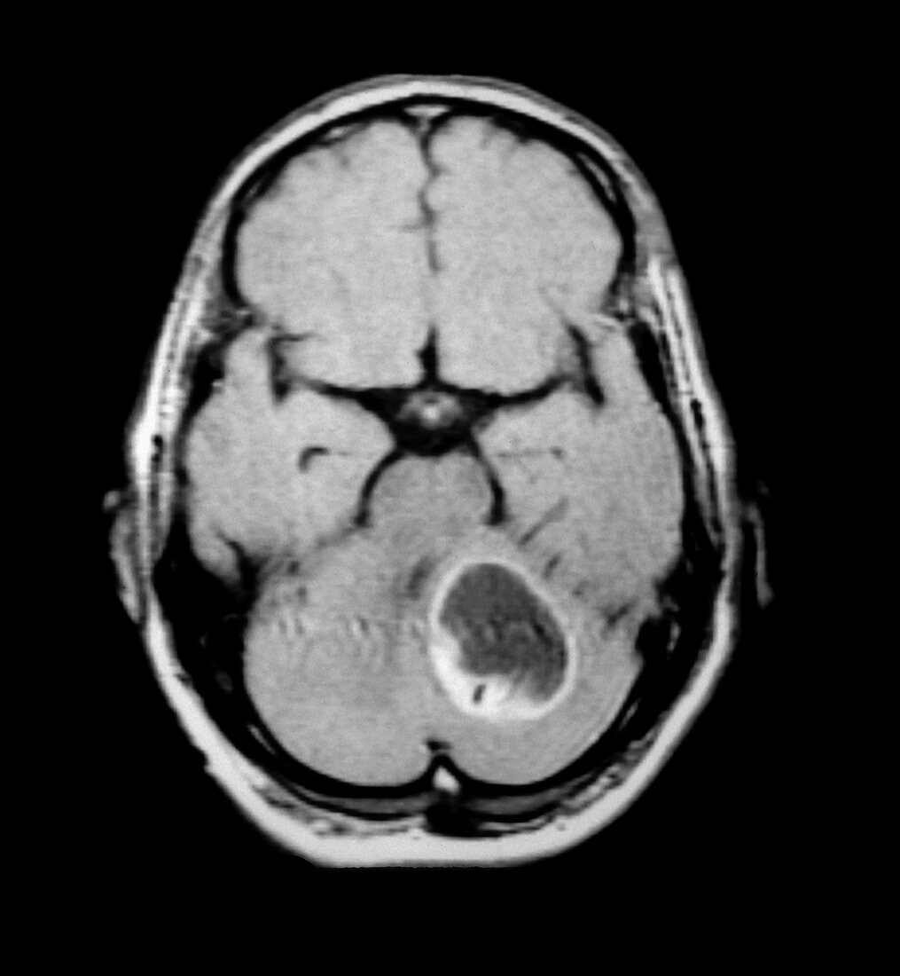
| This contrast enhanced axial (cross sectional) MRI image through the posterior fossa shows a well circumscribed,partially cystic mass with a solid region of pathologic enhancement along the back wall of the mass. This mass is located in the left cerebellum. There is mass effect with compression of the fourth ventricle. This is a typical appearance of a benign pilocytic astrocytoma,which are often found in the pediatric age group. This is one of the few truly benign neoplasms of the brain which when completely resected results in a cure. An astrocytoma is a type of glioma which is the most common type of primary cerebral neoplasm (tumour-). Gliomas arise from the supporting elements in the brain | |
| Licence : | Droits gérés |
| Crédit: | Science Photo Library / Living Art Enterprises, LLC |
| Taille de l’image : | 6716 px × 7280 px |
| Model Release : | Non requis |
| Property Release : | Non requis |
| Restrictions : |
|
Prix pour cette image À partir de 45 €
Produit vendu
(Calendrier, Carte postale, Carte de vœux, Impression sur textile, Packaging etc)
À partir de 45 €
Usage commercial
(Affichage, Annonce presse, Annonce TV, Carte, Digital - hors rés. sociaux, Digital - rés. sociaux etc)
À partir de 45 €
Éditorial
(Digital, Journal, Livre, Livre pratique, Magazine, Télévision etc)
À partir de 60 €
Usage non-commercial
(Digital - hors rés. sociaux, Digital - rés. sociaux etc)
À partir de 120 €