False-col TEM of pancereatic acinar cell from bat
Numéro d’image : 12066087
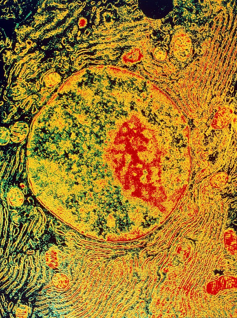
| False-colour transmission electron micrograph of a pancreatic acinar cell from the bat Myotis lucifugus. At center is the nucleus,containing a nucleolus (red). The nucleus is bound by a double membrane separated by a small space,the perinuclear space. Nuclear materials (ions & small molecules) diffuse across the inner membrane into the space & out to the cytoplasm. Nuclear pores (gaps) punctuating the membrane represent a second transport system,conveying larger molecules. Ribosomes adhere to the outer nuclear membrane & to the rough endoplasmic reticulum (parallel layers) in the cytoplasm. They are the sites of cellular protein synthesis. Original B/W print is G455/005 | |
| Licence : | Droits gérés |
| Crédit: | Science Photo Library / Fawcett, Dr. Don |
| Taille de l’image : | 3613 px × 4848 px |
| Model Release : | Non requis |
| Property Release : | Non requis |
| Restrictions : |
|
Prix pour cette image À partir de 45 €
Produit vendu
(Calendrier, Carte postale, Carte de vœux, Impression sur textile, Packaging etc)
À partir de 45 €
Usage commercial
(Affichage, Annonce presse, Annonce TV, Carte, Digital - hors rés. sociaux, Digital - rés. sociaux etc)
À partir de 45 €
Éditorial
(Digital, Journal, Livre, Livre pratique, Magazine, Télévision etc)
À partir de 60 €
Usage non-commercial
(Digital - hors rés. sociaux, Digital - rés. sociaux etc)
À partir de 120 €