Fetal skeleton,light micrograph
Numéro d’image : 12052696
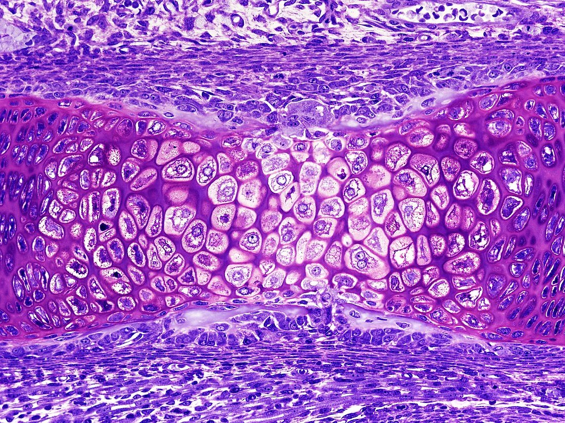
| Light microscopy of a longitudinal section through a fetal finger. At this stage there is no bone tissue but rather a cartilage model acting as a precursor of the bone. All of the cells are chondrocytes. They are programmed to die but their secreted cartilage matrix (purple-red colour) provides the scaffold upon which bone matrix will later be deposited. The replacement of cartilage with bone is referred to as endochondral ossification.Magnification x90 when printed at 10 cm | |
| Licence : | Droits gérés |
| Crédit: | Science Photo Library / Microscape |
| Taille de l’image : | 4961 px × 3720 px |
| Model Release : | Non requis |
| Property Release : | Non requis |
| Restrictions : | - |
Prix pour cette image À partir de 45 €
Produit vendu
(Calendrier, Carte postale, Carte de vœux, Impression sur textile, Packaging etc)
À partir de 45 €
Usage commercial
(Affichage, Annonce presse, Annonce TV, Carte, Digital - hors rés. sociaux, Digital - rés. sociaux etc)
À partir de 45 €
Éditorial
(Digital, Journal, Livre, Livre pratique, Magazine, Télévision etc)
À partir de 60 €
Usage non-commercial
(Digital - hors rés. sociaux, Digital - rés. sociaux etc)
À partir de 120 €