Skeletal muscle,light micrograph
Numéro d’image : 12052695
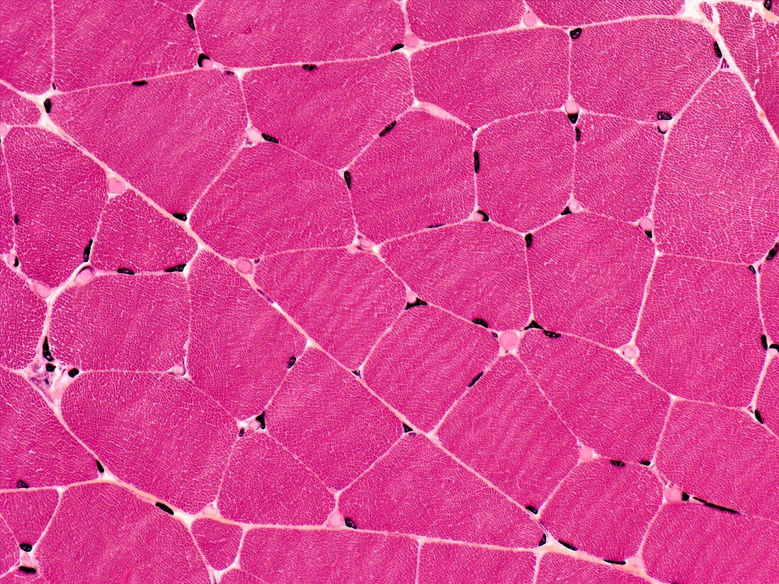
| Light microscopy of skeletal muscle fibres seen in cross section and separated by connective tissue carrying blood vessels and nerves. The stippled appearance is due to many individual units of contractile filaments called myofibrils. The nuclei of each fibre (i.e. a muscle cell) are displaced to the periphery and typical of skeletal muscle. Magnification x250 when printed at 10 cm | |
| Licence : | Droits gérés |
| Crédit: | Science Photo Library / Microscape |
| Taille de l’image : | 4823 px × 3617 px |
| Model Release : | Non requis |
| Property Release : | Non requis |
| Restrictions : | - |
Prix pour cette image À partir de 45 €
Produit vendu
(Calendrier, Carte postale, Carte de vœux, Impression sur textile, Packaging etc)
À partir de 45 €
Usage commercial
(Affichage, Annonce presse, Annonce TV, Carte, Digital - hors rés. sociaux, Digital - rés. sociaux etc)
À partir de 45 €
Éditorial
(Digital, Journal, Livre, Livre pratique, Magazine, Télévision etc)
À partir de 60 €
Usage non-commercial
(Digital - hors rés. sociaux, Digital - rés. sociaux etc)
À partir de 120 €
Mots clés
- aucun,
- biologie,
- biologique,
- catégorie,
- coupe,
- fibre musculaire,
- hématoxyline,
- histologie,
- histologique,
- microscope optique,
- microscope photonique,
- microscopie optique,
- microscopie photonique,
- muscle,
- muscle squelettique,
- muscle strié,
- muscle volontaire,
- myofibrille,
- partie,
- personne,
- section,
- tache d'éosine