TEM of Adipose Cells
Numéro d’image : 12046440
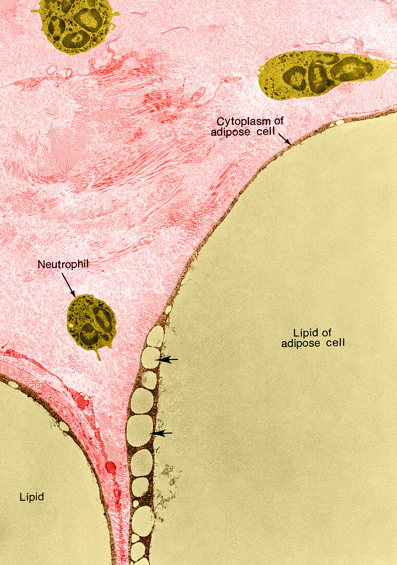
| Colour enhanced transmission electron micrograph of portions of two adipose cells and associated connective tissue from the epididymal fat pad of a rat. Adipose cells are among the largest cells in the body,but less than one fortieth of their total volume is metabolically active. The nucleus is displaced to the periphery and the cytoplasm is reduced to a thin layer surrounding a large globule of accumulated lipid. The very large relative size of adipose cells can be appreciated in the accompanying micrograph by comparison of three leucocytes in the field with portions of two fat cells included in the lower half of the figure. Newly synthesized lipid first forms small droplets in the peripheral layer of cytoplasm (at arrows) and these subsequently coalesce with the large central lipid drop | |
| Licence : | Droits gérés |
| Crédit: | Science Photo Library / Fawcett, Don W. |
| Taille de l’image : | 3347 px × 4762 px |
| Model Release : | Non requis |
| Property Release : | Non requis |
| Restrictions : |
|
Prix pour cette image À partir de 45 €
Produit vendu
(Calendrier, Carte postale, Carte de vœux, Impression sur textile, Packaging etc)
À partir de 45 €
Usage commercial
(Affichage, Annonce presse, Annonce TV, Carte, Digital - hors rés. sociaux, Digital - rés. sociaux etc)
À partir de 45 €
Éditorial
(Digital, Journal, Livre, Livre pratique, Magazine, Télévision etc)
À partir de 60 €
Usage non-commercial
(Digital - hors rés. sociaux, Digital - rés. sociaux etc)
À partir de 120 €
Mots clés
- adipeux,
- adipocyte,
- amélioré,
- aucun,
- augmenté,
- cellule,
- cellule de rat,
- cellule rat,
- colorié,
- colorisé,
- cytoplasme,
- histologie,
- Leucocyte,
- lipidique,
- M.E.T.,
- MET,
- micrographie,
- microscope,
- microscope électronique à transmission,
- microscope électronique en transmission,
- microscopie,
- neutrophile,
- personne,
- renforcé,
- système reproductif