Meiosis after Radiation Exposure
Numéro d’image : 12044587
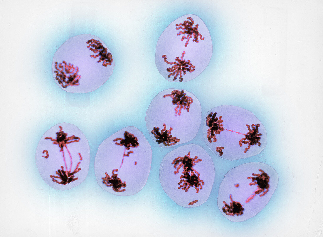
| Colour-enhanced micrograph of cells of Trillium erectum in division (meiotic first anaphase). The cells were exposed to an acute dose of 50 R of X-rays at early prophase. The three cells at the top and centre show no signs of chromosome damage. The other cells contain bridges between chromosomes and fragments of chromosomes. Magnification unknown. This image has been colour-enhanced | |
| Licence : | Droits gérés |
| Crédit: | Science Photo Library / Brookhaven National Laboratory |
| Taille de l’image : | 4138 px × 3047 px |
| Model Release : | Non requis |
| Property Release : | Non requis |
| Restrictions : |
|
Prix pour cette image À partir de 45 €
Produit vendu
(Calendrier, Carte postale, Carte de vœux, Impression sur textile, Packaging etc)
À partir de 45 €
Usage commercial
(Affichage, Annonce presse, Annonce TV, Carte, Digital - hors rés. sociaux, Digital - rés. sociaux etc)
À partir de 45 €
Éditorial
(Digital, Journal, Livre, Livre pratique, Magazine, Télévision etc)
À partir de 60 €
Usage non-commercial
(Digital - hors rés. sociaux, Digital - rés. sociaux etc)
À partir de 120 €
Mots clés
- anaphase,
- biologie,
- botanique,
- chromosome,
- coloration,
- colorié,
- colorisation,
- colorisé,
- couleur,
- couleur améliorée,
- couleur augmentée,
- couleurs améliorées,
- division,
- division cellulaire,
- gène,
- génétique,
- méiose,
- microbiologie,
- micrographie,
- microscope,
- microscopie,
- nucléaire,
- rayonnement,
- rayons X,
- science,
- séparation,
- trillium,
- trillium erectum