Epidural Space
Numéro d’image : 12042518
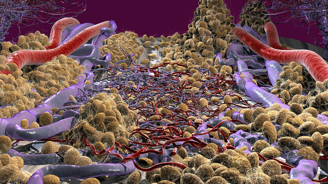
| This microscopic view of the epidural space demonstrates the dense venous plexus and capillaries present throughout this space. Fat cells (yellow) are seen scattered throughout the space connected by connective tissue. The posterior longitudinal ligament is barely visible in the background under fat cells at the top of the image | |
| Licence : | Droits gérés |
| Crédit: | Science Photo Library / Orthoclick |
| Taille de l’image : | 4552 px × 2561 px |
| Model Release : | Non requis |
| Property Release : | Non requis |
| Restrictions : |
|
Prix pour cette image À partir de 45 €
Produit vendu
(Calendrier, Carte postale, Carte de vœux, Impression sur textile, Packaging etc)
À partir de 45 €
Usage commercial
(Affichage, Annonce presse, Annonce TV, Carte, Digital - hors rés. sociaux, Digital - rés. sociaux etc)
À partir de 45 €
Éditorial
(Digital, Journal, Livre, Livre pratique, Magazine, Télévision etc)
À partir de 60 €
Usage non-commercial
(Digital - hors rés. sociaux, Digital - rés. sociaux etc)
À partir de 120 €