Breast Cancer Stages,Illustration
Numéro d’image : 12041641
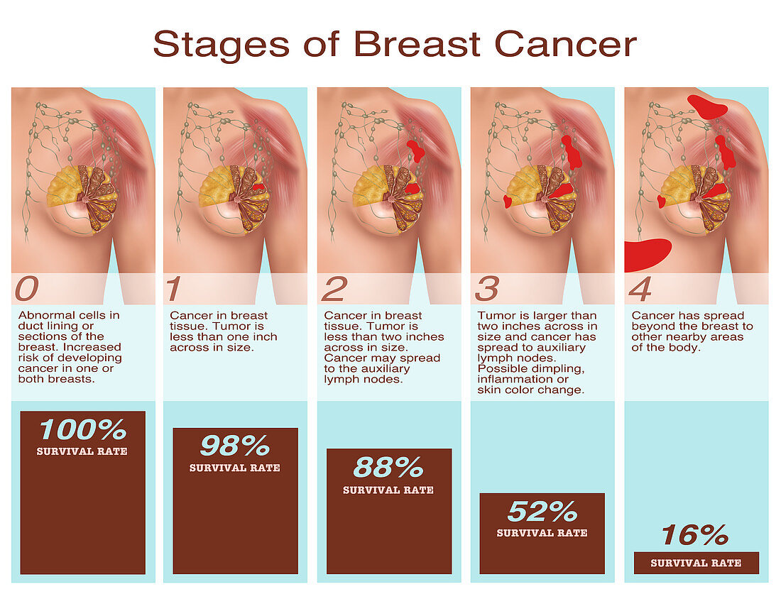
| Illustration showing stages of breast cancer and the survival rate at each stage. Starting at the left image 0 shows abnormal cells in duct lining or sections of the breast giving an increased risk of developing cancer in one or both breasts. Image 1 shows cancer in breast tissue,with the tumour being less than 1inch across in size. The 2nd image shows a tumour less than 2 inches wide,and a possibility of cancer spreading to the axillary lymph nodes. Image 3 shows the tumour measuring larger than two inches across in size and cancer has spread to axillary lymph nodes. The final image shows that cancer has spread beyond the breast to other nearby areas of the body | |
| Licence : | Droits gérés |
| Crédit: | Science Photo Library / Shockey, Gwen |
| Taille de l’image : | 5520 px × 4200 px |
| Model Release : | Non requis |
| Property Release : | Non requis |
| Restrictions : |
|
Prix pour cette image À partir de 45 €
Produit vendu
(Calendrier, Carte postale, Carte de vœux, Impression sur textile, Packaging etc)
À partir de 45 €
Usage commercial
(Affichage, Annonce presse, Annonce TV, Carte, Digital - hors rés. sociaux, Digital - rés. sociaux etc)
À partir de 45 €
Éditorial
(Digital, Journal, Livre, Livre pratique, Magazine, Télévision etc)
À partir de 60 €
Usage non-commercial
(Digital - hors rés. sociaux, Digital - rés. sociaux etc)
À partir de 120 €
Mots clés
- anatomie,
- anatomique,
- annoté,
- cancer,
- cancer du sein,
- cancéreux,
- cellules anormales,
- étapes,
- féminin,
- féminine,
- ganglions lymphatiques axillaires,
- graphique d'information,
- graphique informatif,
- illustration,
- infographie,
- infographique,
- information graphique,
- maladie,
- malin,
- oeuvre,
- oncologie,
- pathologique,
- poitrine,
- progression du cancer,
- propagation du cancer,
- sein,
- tissu de la poitrine,
- tissu du sein,
- tumeur