Breast Anatomy,Illustration
Numéro d’image : 12041635
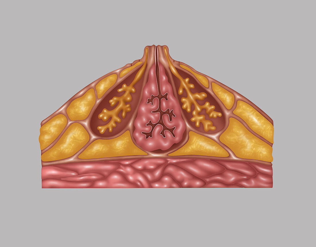
| Illustration detailing the anatomy of a female breast from a side view. The image shows the following: montgomery's gland and superficial fascia (outer lining),subcutaneous fat (orange outer sections),ampulla and lactiferous duct and connective tissue (pink centre),coopers ligaments,mammary fat (orange bottom sections),pectoral fascia,and pectoralis major (pink at bottom) | |
| Licence : | Droits gérés |
| Crédit: | Science Photo Library / Shockey, Gwen |
| Taille de l’image : | 3966 px × 3102 px |
| Model Release : | Non requis |
| Property Release : | Non requis |
| Restrictions : |
|
Prix pour cette image À partir de 45 €
Produit vendu
(Calendrier, Carte postale, Carte de vœux, Impression sur textile, Packaging etc)
À partir de 45 €
Usage commercial
(Affichage, Annonce presse, Annonce TV, Carte, Digital - hors rés. sociaux, Digital - rés. sociaux etc)
À partir de 45 €
Éditorial
(Digital, Journal, Livre, Livre pratique, Magazine, Télévision etc)
À partir de 60 €
Usage non-commercial
(Digital - hors rés. sociaux, Digital - rés. sociaux etc)
À partir de 120 €