Lingual Thyroid,CT Scan
Numéro d’image : 12036566
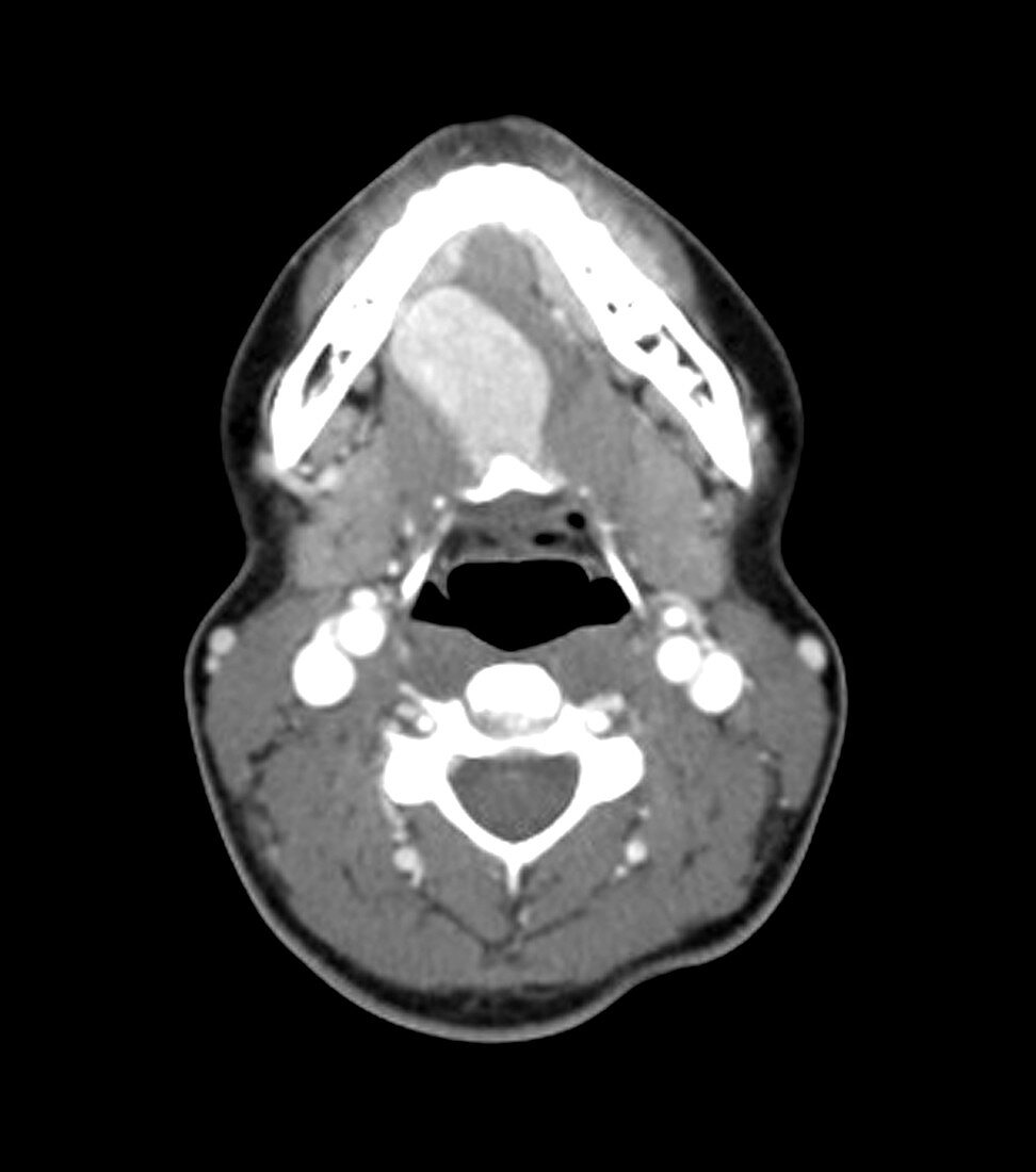
| This contrast-enhanced,axial (cross-sectional) CT image through the oral cavity shows a large,high-density (looks white) region in the tongue. This represents lingual thyroid tissue. This patient had no thyroid tissue in the expected region of the lower neck. The thyroid gland develops near the foramen cecum of the tongue and normally descends along a tract called the thyroglossal duct to the lower neck. Rarely,this normal descent does not occur and thyroid tissue remains in the tongue,but remnants of thyroid tissue and the duct itself may occur anywhere along the tract. Often,however,no thyroid tissue is present in the lower neck,only in the tongue base,as in this case | |
| Licence : | Droits gérés |
| Crédit: | Science Photo Library / Living Art Enterprises |
| Taille de l’image : | 4200 px × 4755 px |
| Model Release : | Non requis |
| Property Release : | Non requis |
| Restrictions : |
|
Prix pour cette image À partir de 45 €
Produit vendu
(Calendrier, Carte postale, Carte de vœux, Impression sur textile, Packaging etc)
À partir de 45 €
Usage commercial
(Affichage, Annonce presse, Annonce TV, Carte, Digital - hors rés. sociaux, Digital - rés. sociaux etc)
À partir de 45 €
Éditorial
(Digital, Journal, Livre, Livre pratique, Magazine, Télévision etc)
À partir de 60 €
Usage non-commercial
(Digital - hors rés. sociaux, Digital - rés. sociaux etc)
À partir de 120 €
Mots clés
- anormal,
- caecum,
- canal,
- cecum,
- conduit,
- désordre,
- diagnostic,
- diagnostic par imagerie,
- diagnostique,
- diagnostique par imagerie,
- état,
- FORAMEN,
- FORAMEN CECUM,
- glande,
- glande thyroïde,
- imagerie médicale,
- kyste,
- langue,
- lésion,
- lingual,
- maladie,
- malsain,
- masse,
- médecine,
- médical,
- médicale,
- pathologie,
- représentation,
- science,
- thyroïde,
- trouble,
- vestige