Carotid Cavernous Sinus Fistula,MRI
Numéro d’image : 12036543
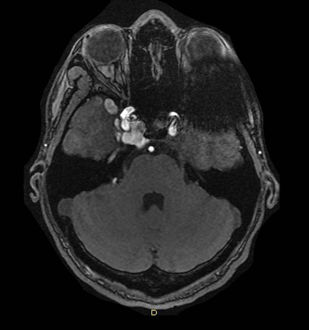
| This axial source image from an intracranial MRA demonstrates pathologic enlargement of the cavernous sinus (on the viewer's left),in addition to abnormal enlarged venous channels (white and grey) in the anterior middle cranial fossa. These changes reflect a post-traumatic carotid cavernous sinus fistula. These occur as a result of a traumatic tear of the internal carotid artery within the cavernous sinus,resulting in an arterial-venous shunt directly into the cavernous sinus and its venous outflow,which often includes orbital venous drainage. The degree of cortical venous drainage/congestion directly correlates with the biologic aggressiveness of these lesions. Those with abundant cortical venous drainage/outflow have a high risk for intracranial hemorrhage. These are often treated through endovascular embolization techniques | |
| Licence : | Droits gérés |
| Crédit: | Science Photo Library / Living Art Enterprises |
| Taille de l’image : | 4200 px × 4482 px |
| Model Release : | Non requis |
| Property Release : | Non requis |
| Restrictions : |
|
Prix pour cette image À partir de 45 €
Produit vendu
(Calendrier, Carte postale, Carte de vœux, Impression sur textile, Packaging etc)
À partir de 45 €
Usage commercial
(Affichage, Annonce presse, Annonce TV, Carte, Digital - hors rés. sociaux, Digital - rés. sociaux etc)
À partir de 45 €
Éditorial
(Digital, Journal, Livre, Livre pratique, Magazine, Télévision etc)
À partir de 60 €
Usage non-commercial
(Digital - hors rés. sociaux, Digital - rés. sociaux etc)
À partir de 120 €
Mots clés
- anormal,
- arracher,
- artère carotide,
- artère carotide interne,
- blessé,
- blessure,
- carotide,
- CC,
- déchirer,
- désordre,
- diagnostic,
- diagnostic par imagerie,
- diagnostique,
- diagnostique par imagerie,
- état,
- fistule,
- I.R.M.,
- imagerie médicale,
- imagerie par résonnance magnétique,
- IRM,
- larme,
- M.,
- maladie,
- malsain,
- médecine,
- médical,
- médicale,
- pathologie,
- représentation,
- science,
- sinus,
- sinus caverneux,
- trauma,
- traumatique,
- traumatisme,
- trouble