Enhanced Large Meningioma on MRI
Numéro d’image : 12036399
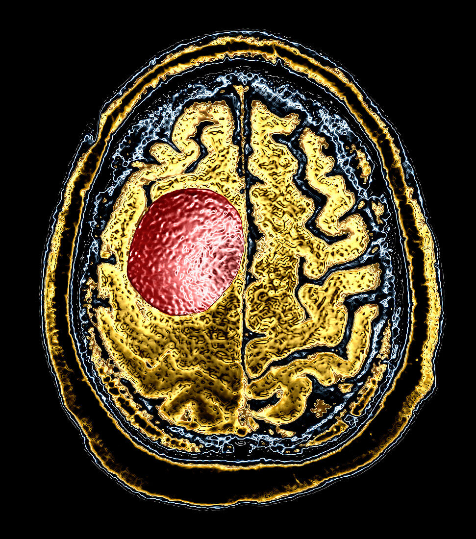
| This colour enhanced axial (cross sectional) T1-weighted MRI image of the brain shows a large,well circumscribed round mass (red) in the high posterior frontal lobe in the supplementary motor/pre-motor region compatible with a meningioma on viewers left. These account for about 20% of all intracranial masses in adults and are generally benign lesions however a few may be aggressive,infiltrating adjacent brain and growing rapidly. Occasionally they are multiple in a single patient | |
| Licence : | Droits gérés |
| Crédit: | Science Photo Library / Living Art Enterprises, LLC |
| Taille de l’image : | 4200 px × 4757 px |
| Model Release : | Non requis |
| Property Release : | Non requis |
| Restrictions : |
|
Prix pour cette image À partir de 45 €
Produit vendu
(Calendrier, Carte postale, Carte de vœux, Impression sur textile, Packaging etc)
À partir de 45 €
Usage commercial
(Affichage, Annonce presse, Annonce TV, Carte, Digital - hors rés. sociaux, Digital - rés. sociaux etc)
À partir de 45 €
Éditorial
(Digital, Journal, Livre, Livre pratique, Magazine, Télévision etc)
À partir de 60 €
Usage non-commercial
(Digital - hors rés. sociaux, Digital - rés. sociaux etc)
À partir de 120 €
Mots clés
- anormal,
- cerveau,
- I.R.M.,
- imagerie par résonance magnétique,
- imagerie par résonnance magnétique,
- IRM,
- IRM du cerveau,
- masse cérébrale,
- masse du cerveau,
- méningiome,
- neurofibromatose type 2,
- neurofibromatose type II,
- NF II,
- T1,
- T1-pondéré,
- tumeur,
- tumeur cérébrale bénigne,
- tumeur intra-crânienne,
- tumeur intracrânienne,
- tumeurs cérébrales