Cholesteatoma of Temporal Bone
Numéro d’image : 12036369
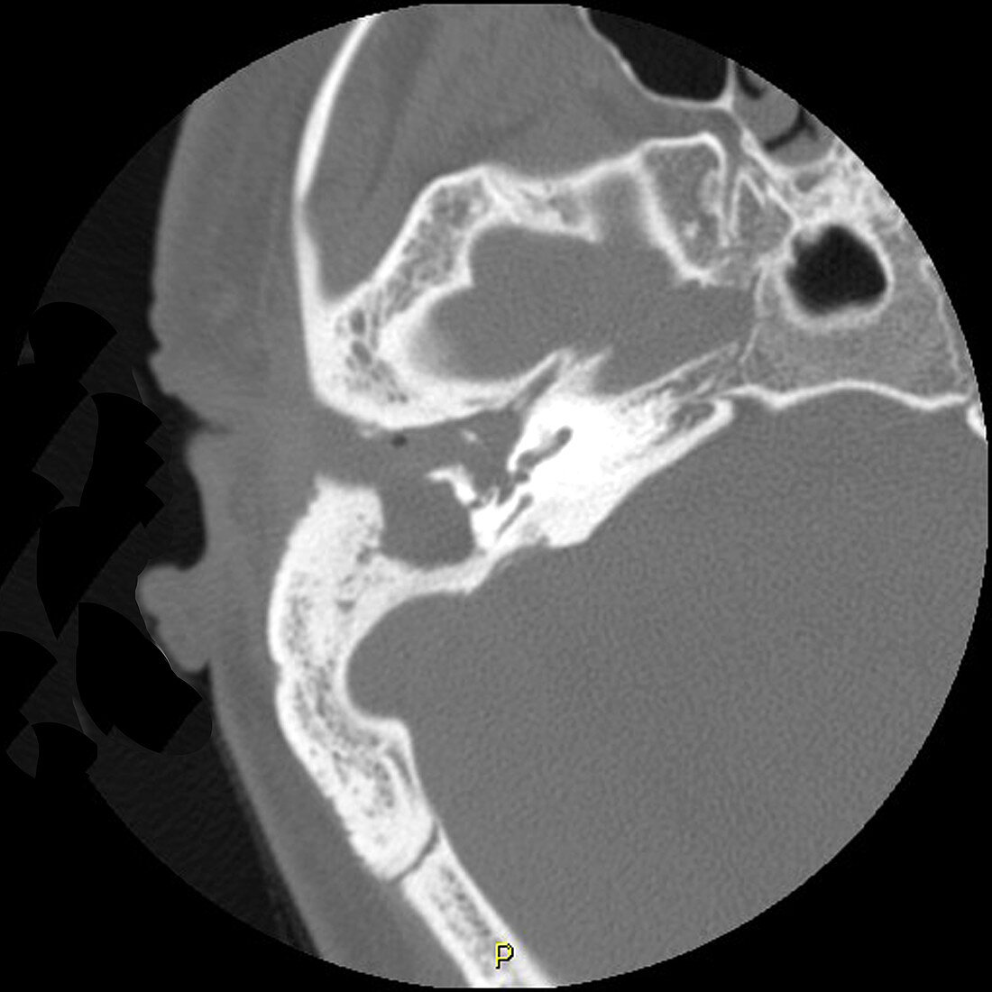
| This high resolution thin section axial (cross sectional) CT image of the temporal bone shows a destructive soft tissue lesion in the middle ear (tympanic cavity) and mastoid antrum. There is also opacification of the right external auditory canal. The ossicles (small bones in the middle ear) have been largely destroyed. Cholesteatomas are usually the result of a perforation (tear) of the tympanic membrane (ear drum) with ingrowth of squamous epithelial cells which shed desquamated cellular debris and keratin. However they can be congenital .These are benign lesions which progressively enlarge and are locally destructive | |
| Licence : | Droits gérés |
| Crédit: | Science Photo Library / Living Art Enterprises, LLC |
| Taille de l’image : | 4200 px × 4200 px |
| Model Release : | Non requis |
| Property Release : | Non requis |
| Restrictions : |
|
Prix pour cette image À partir de 45 €
Produit vendu
(Calendrier, Carte postale, Carte de vœux, Impression sur textile, Packaging etc)
À partir de 45 €
Usage commercial
(Affichage, Annonce presse, Annonce TV, Carte, Digital - hors rés. sociaux, Digital - rés. sociaux etc)
À partir de 45 €
Éditorial
(Digital, Journal, Livre, Livre pratique, Magazine, Télévision etc)
À partir de 60 €
Usage non-commercial
(Digital - hors rés. sociaux, Digital - rés. sociaux etc)
À partir de 120 €