Neonatal Transcranial Ultrasound
Numéro d’image : 12036362
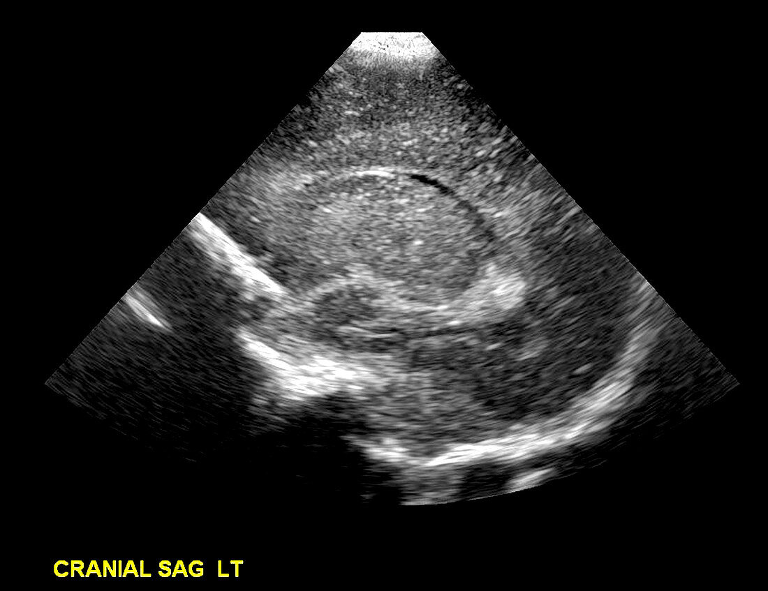
| This sagital (from the side) ultrasound image obtained through the anterior fontanella shows the normal appearance of the lateral ventricle and the choroid plexus within the atrium of the lateral ventricle. The thalamus is well seen between the body and temporal horns of the lateral ventricle. The ventricles are not enlarged (no hydrocephalus). The gaps present within the fetal skull (fontanelles) are used as acoustic windows to view the intracranial structures. Ultrasound is commonly and frequently used to image the newborn and young infant because of its ease (machine can be transported to the neonatal ICA easily) and there is no ionizing radiation | |
| Licence : | Droits gérés |
| Crédit: | Science Photo Library / Living Art Enterprises, LLC |
| Taille de l’image : | 5454 px × 4200 px |
| Model Release : | Non requis |
| Property Release : | Non requis |
| Restrictions : |
|
Prix pour cette image À partir de 45 €
Produit vendu
(Calendrier, Carte postale, Carte de vœux, Impression sur textile, Packaging etc)
À partir de 45 €
Usage commercial
(Affichage, Annonce presse, Annonce TV, Carte, Digital - hors rés. sociaux, Digital - rés. sociaux etc)
À partir de 45 €
Éditorial
(Digital, Journal, Livre, Livre pratique, Magazine, Télévision etc)
À partir de 60 €
Usage non-commercial
(Digital - hors rés. sociaux, Digital - rés. sociaux etc)
À partir de 120 €