Cervical Myelogram CT
Numéro d’image : 12036353
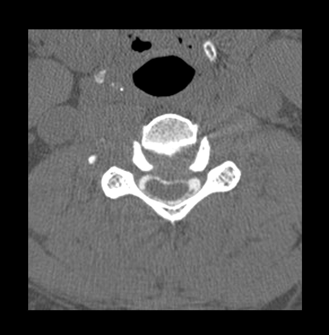
| This axial (cross sectional) CT image of the cervical spine was performed after contrast was injected into the lumbar subarachnoid space via a spinal tap. The contrast (white) outlines the spinal cord (ovoid grey structure) and aids in delineating disc herniation and the possible mass effect they may have on nerve roots and the spinal cord. This person has a congenitally small spinal canal in addition to degenerative changes with multiple disc herniations. A disc herniation (grey material compressing the white contrast above the spinal cord) at this level is compressing and deforming the spinal cord | |
| Licence : | Droits gérés |
| Crédit: | Science Photo Library / Living Art Enterprises, LLC |
| Taille de l’image : | 4200 px × 4272 px |
| Model Release : | Non requis |
| Property Release : | Non requis |
| Restrictions : |
|
Prix pour cette image À partir de 45 €
Produit vendu
(Calendrier, Carte postale, Carte de vœux, Impression sur textile, Packaging etc)
À partir de 45 €
Usage commercial
(Affichage, Annonce presse, Annonce TV, Carte, Digital - hors rés. sociaux, Digital - rés. sociaux etc)
À partir de 45 €
Éditorial
(Digital, Journal, Livre, Livre pratique, Magazine, Télévision etc)
À partir de 60 €
Usage non-commercial
(Digital - hors rés. sociaux, Digital - rés. sociaux etc)
À partir de 120 €