Enhanced Cerebral Cavernous Malformation
Numéro d’image : 12036336
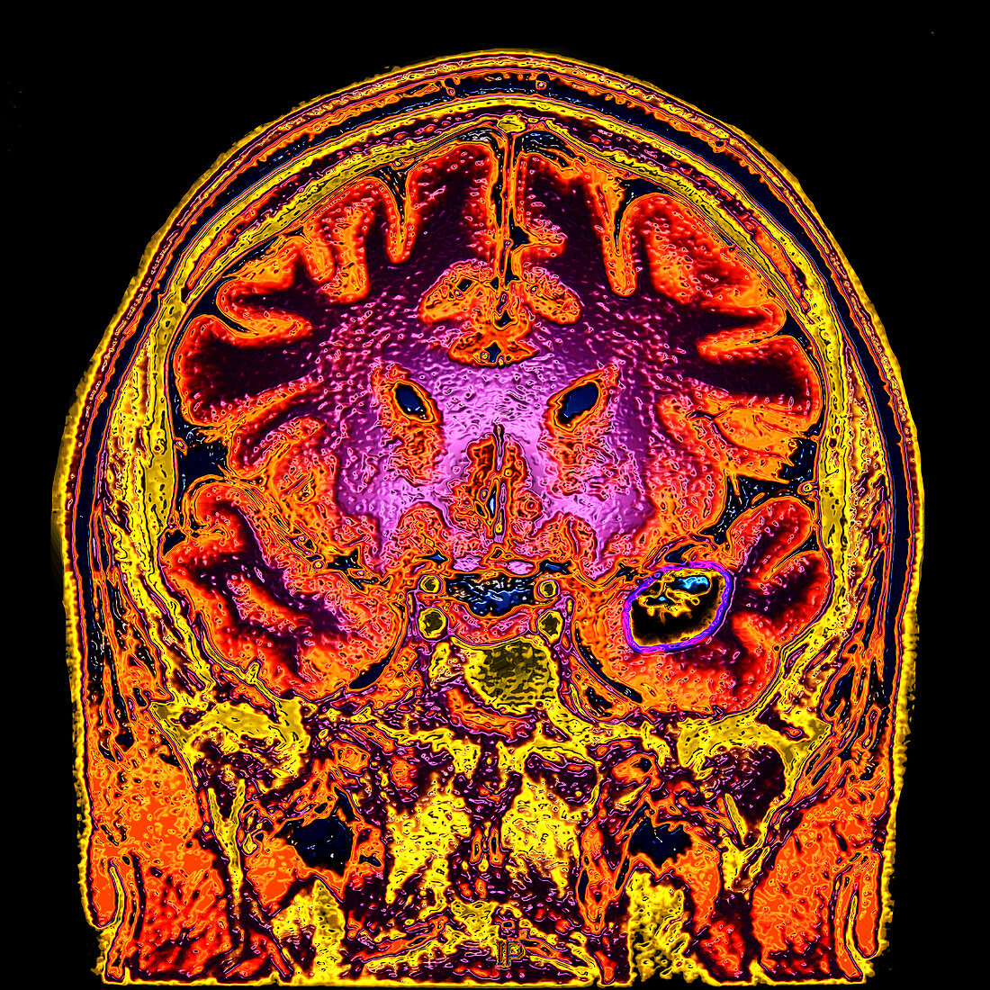
| This colour enhanced coronal (frontal view) T2-weighted MRI image shows the typical appearance of a cavernous malformation in the temporal lobe (on your right). These are slow flow vascular malformations that contain dilated vascular spaces without any intervening brain. The can be inherited (more common) or acquired. They can occur anywhere and symptoms depend on upon their location and if repeated hemorrhage occur | |
| Licence : | Droits gérés |
| Crédit: | Science Photo Library / Living Art Enterprises, LLC |
| Taille de l’image : | 4200 px × 4200 px |
| Model Release : | Non requis |
| Property Release : | Non requis |
| Restrictions : |
|
Prix pour cette image À partir de 45 €
Produit vendu
(Calendrier, Carte postale, Carte de vœux, Impression sur textile, Packaging etc)
À partir de 45 €
Usage commercial
(Affichage, Annonce presse, Annonce TV, Carte, Digital - hors rés. sociaux, Digital - rés. sociaux etc)
À partir de 45 €
Éditorial
(Digital, Journal, Livre, Livre pratique, Magazine, Télévision etc)
À partir de 60 €
Usage non-commercial
(Digital - hors rés. sociaux, Digital - rés. sociaux etc)
À partir de 120 €