Temporal Lobe AVM on MRI
Numéro d’image : 12036296
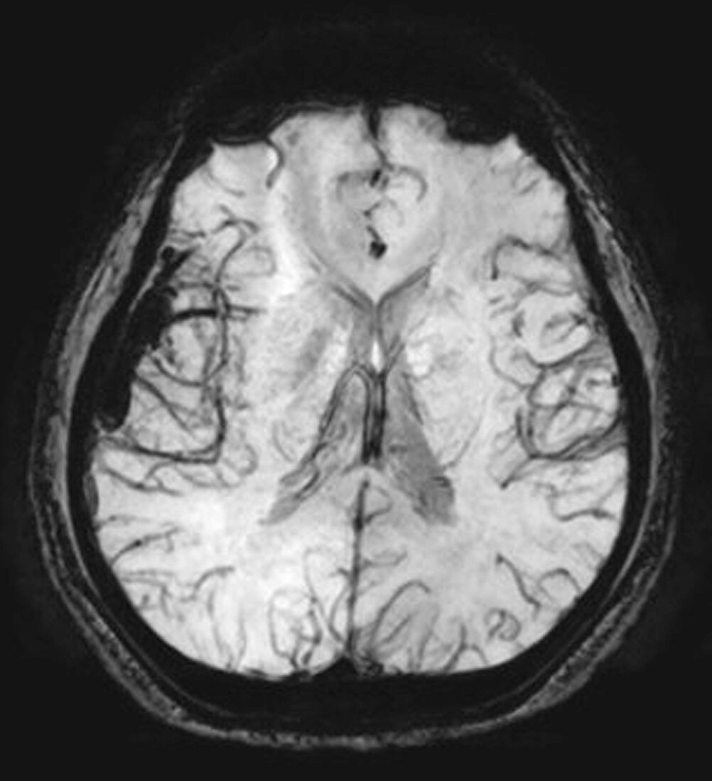
| This axial (cross sectional) susceptibility-weighted MR image (SWI) shows multiple small regions of dark signal on the viewers left in the temporal lobe representing multiple abnormal small blood vessels of a high flow arterial venous malformation (AVM). There is a single large curvilinear region of dark signal representing a large vein draining the AVM. AVMs are dangerous because of there potential for hemorrhage (bleeding) but they can also result in strokes,seizures,headaches and even death | |
| Licence : | Droits gérés |
| Crédit: | Science Photo Library / Living Art Enterprises, LLC |
| Taille de l’image : | 4200 px × 4609 px |
| Model Release : | Non requis |
| Property Release : | Non requis |
| Restrictions : |
|
Prix pour cette image À partir de 45 €
Produit vendu
(Calendrier, Carte postale, Carte de vœux, Impression sur textile, Packaging etc)
À partir de 45 €
Usage commercial
(Affichage, Annonce presse, Annonce TV, Carte, Digital - hors rés. sociaux, Digital - rés. sociaux etc)
À partir de 45 €
Éditorial
(Digital, Journal, Livre, Livre pratique, Magazine, Télévision etc)
À partir de 60 €
Usage non-commercial
(Digital - hors rés. sociaux, Digital - rés. sociaux etc)
À partir de 120 €
Mots clés
- anormal,
- cerveau,
- hémorragie cérébrale,
- hémorragie intracrânienne,
- I.R.M.,
- image ponderée fonction sensibilité,
- imagerie par résonance magnétique,
- imagerie par résonnance magnétique,
- IRM,
- lésion,
- lésion vasculaire,
- malformation artérioveineuse,
- malformation artéro-veineuse,
- masse cérébrale,
- masse du cerveau,
- saignement cérébral,
- shunt vasculaire,
- vaisseaux sanguins cérébraux