Enhanced Haemorrhagic Arachnoid Cyst
Numéro d’image : 12036285
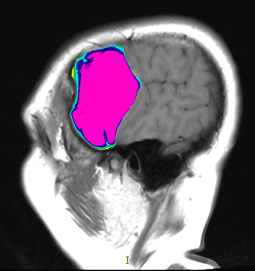
| This colour enhanced sagittal (from the side) T1-weighted MRI image of the brain without contrast demonstrates the typical appearance of an arachnoid cyst in the middle cranial fossa however this person sustained a head injury resulting in hemorrhage/bleeding into this extra-axial cyst. Deformity/dysmorphic anterior temporal lobe adjacent to the arachnoid cyst would be expected whether there had or had not been bleeding into the cyst | |
| Licence : | Droits gérés |
| Crédit: | Science Photo Library / Living Art Enterprises, LLC |
| Taille de l’image : | 4200 px × 4456 px |
| Model Release : | Non requis |
| Property Release : | Non requis |
| Restrictions : |
|
Prix pour cette image À partir de 45 €
Produit vendu
(Calendrier, Carte postale, Carte de vœux, Impression sur textile, Packaging etc)
À partir de 45 €
Usage commercial
(Affichage, Annonce presse, Annonce TV, Carte, Digital - hors rés. sociaux, Digital - rés. sociaux etc)
À partir de 45 €
Éditorial
(Digital, Journal, Livre, Livre pratique, Magazine, Télévision etc)
À partir de 60 €
Usage non-commercial
(Digital - hors rés. sociaux, Digital - rés. sociaux etc)
À partir de 120 €