Meningitis in Newborn
Numéro d’image : 12036265
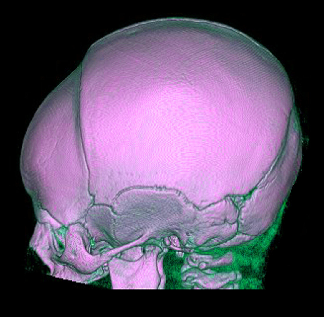
| This lateral (from the side) 3D CT reconstruction of the skull in this infant several months after surviving meningitis shows evidence of severe brain atrophy (loss of brain tissue) as a complication of the meninigitis. Due to the low volume of the brain the skull is not expanding as it would normally do. Instead the frontal bone has collapsed due to the lack of normal expansion by the growing brain. This appearance of a small brain and skull is called microcephaly | |
| Licence : | Droits gérés |
| Crédit: | Science Photo Library / Living Art Enterprises, LLC |
| Taille de l’image : | 4292 px × 4200 px |
| Model Release : | Non requis |
| Property Release : | Non requis |
| Restrictions : |
|
Prix pour cette image À partir de 45 €
Produit vendu
(Calendrier, Carte postale, Carte de vœux, Impression sur textile, Packaging etc)
À partir de 45 €
Usage commercial
(Affichage, Annonce presse, Annonce TV, Carte, Digital - hors rés. sociaux, Digital - rés. sociaux etc)
À partir de 45 €
Éditorial
(Digital, Journal, Livre, Livre pratique, Magazine, Télévision etc)
À partir de 60 €
Usage non-commercial
(Digital - hors rés. sociaux, Digital - rés. sociaux etc)
À partir de 120 €