CT Reconstruction of Intracranial Shunt
Numéro d’image : 12036253
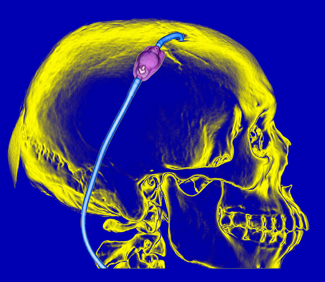
| This colour enhanced lateral (from the side) 3D CT reconstruction of the skull beautifully demonstrates the normal appearance of a ventricular shunt catheter exiting the skull through a frontal burr hole. Over the surface of the skull you see the shunt catheter reservoir (purple) and the shunt tubing (blue) extending inferiorly within the soft tissues of the scalp. These often terminate in the peritoneal cavity of the abdomen or the pleural space of the chest. There are no breaks or discontinuities of the catheter which one would look for with shunt malfunction | |
| Licence : | Droits gérés |
| Crédit: | Science Photo Library / Living Art Enterprises, LLC |
| Taille de l’image : | 4837 px × 4200 px |
| Model Release : | Non requis |
| Property Release : | Non requis |
| Restrictions : |
|
Prix pour cette image À partir de 45 €
Produit vendu
(Calendrier, Carte postale, Carte de vœux, Impression sur textile, Packaging etc)
À partir de 45 €
Usage commercial
(Affichage, Annonce presse, Annonce TV, Carte, Digital - hors rés. sociaux, Digital - rés. sociaux etc)
À partir de 45 €
Éditorial
(Digital, Journal, Livre, Livre pratique, Magazine, Télévision etc)
À partir de 60 €
Usage non-commercial
(Digital - hors rés. sociaux, Digital - rés. sociaux etc)
À partir de 120 €