Brain Activity during Language Task
Numéro d’image : 12035643
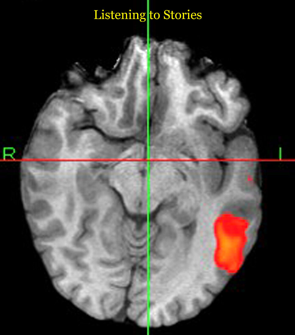
| This axial (cross sectional) T1 weighted MRI image of the brain with colour overlay shows the area of brain activation during a language task. In this task the patient listens passively to stories while special MRI scans are performed. The scans can detect tiny amounts of increased blood flow to the regions of the brain which are activated during the task. The areas of expected brain activation in this case would be Wernicke's Areas (site for language comprehension) which is in the superior temporal gyrus which is the large orange region on the readers right. View 1 of 2 | |
| Licence : | Droits gérés |
| Crédit: | Science Photo Library / Living Art Enterprises |
| Taille de l’image : | 4200 px × 4774 px |
| Model Release : | Non requis |
| Property Release : | Non requis |
| Restrictions : |
|
Prix pour cette image À partir de 45 €
Produit vendu
(Calendrier, Carte postale, Carte de vœux, Impression sur textile, Packaging etc)
À partir de 45 €
Usage commercial
(Affichage, Annonce presse, Annonce TV, Carte, Digital - hors rés. sociaux, Digital - rés. sociaux etc)
À partir de 45 €
Éditorial
(Digital, Journal, Livre, Livre pratique, Magazine, Télévision etc)
À partir de 60 €
Usage non-commercial
(Digital - hors rés. sociaux, Digital - rés. sociaux etc)
À partir de 120 €
Mots clés
- activité cérébrale,
- cerveau,
- cortex cérébral,
- fmri,
- I.R.M.,
- imagerie médicale,
- imagerie par résonance magnétique,
- imagerie par résonnance magnétique,
- IRM,
- IRM fonctionnelle,
- lobe temporal,
- neuro-imagerie,
- neuroimagerie,
- neurologie,
- neurologique,
- région Wernicke,
- scanner du cerveau,
- secteur de Wernicke,
- secteur Wernicke,
- séquence,
- test,
- tester