Submandibular Gland Abscess (CT Scan)
Numéro d’image : 12035633
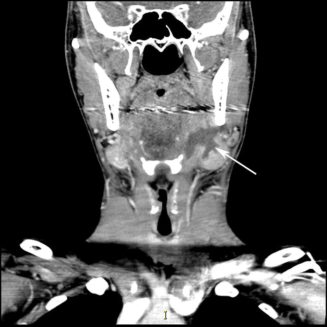
| This axial (cross sectional) contrast enhanced CT image through the upper neck/oral cavity shows the typical appearance of an abscess within the left (on your right) submandibular gland (arrow) extending into the submandibular duct. This is often the result of an obstructing stone in the duct (not in this case) or can be a result of seeding of the gland secondary to infected blood | |
| Licence : | Droits gérés |
| Crédit: | Science Photo Library / Living Art Enterprises |
| Taille de l’image : | 4200 px × 4200 px |
| Model Release : | Non requis |
| Property Release : | Non requis |
| Restrictions : |
|
Prix pour cette image À partir de 45 €
Produit vendu
(Calendrier, Carte postale, Carte de vœux, Impression sur textile, Packaging etc)
À partir de 45 €
Usage commercial
(Affichage, Annonce presse, Annonce TV, Carte, Digital - hors rés. sociaux, Digital - rés. sociaux etc)
À partir de 45 €
Éditorial
(Digital, Journal, Livre, Livre pratique, Magazine, Télévision etc)
À partir de 60 €
Usage non-commercial
(Digital - hors rés. sociaux, Digital - rés. sociaux etc)
À partir de 120 €
Mots clés
- abcès,
- agrandi,
- anomalie,
- anormal,
- anormale,
- anormalité,
- cou,
- diagnostic,
- diagnostique,
- enflée,
- glande,
- glande salivaire,
- glande sous maxillaire,
- glande sous-maxillaire,
- gonflée,
- gonflement,
- infecté,
- infection,
- malsain,
- médical,
- médicale,
- représentation,
- scanner,
- science,
- sous-maxillaire,
- tête,
- tomodensitogramme,
- tomodensitométrie,
- tomographie,
- tomographie assistée par ordinateur