Cervical Myelogram (X-ray)
Numéro d’image : 12035626
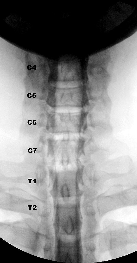
| This frontal X-ray view of the neck demonstrates what a cervical myelogram looks like. This procedure is being done less frequently because of the widespread utilization of MRI,however they still are being used for instances when patients with pacemakers can't have an MRI or when the findings on the MRI are inconclusive. This test is obtained by first doing a spinal tap in the lower back (lumbar) region. Then a contrast agent (dye) is injected where the spinal fluid is located. When the contrast is seen in the neck,X-rays are obtained. This view shows deformity of some of the nerve roots sleeves which normally fill with the dye. The deformities are seen as incomplete filling and in some cases complete lack of filling of the root sleeves with the dye. The various spinal levels are annotated | |
| Licence : | Droits gérés |
| Crédit: | Science Photo Library / Living Art Enterprises |
| Taille de l’image : | 4200 px × 8058 px |
| Model Release : | Non requis |
| Property Release : | Non requis |
| Restrictions : |
|
Prix pour cette image À partir de 45 €
Produit vendu
(Calendrier, Carte postale, Carte de vœux, Impression sur textile, Packaging etc)
À partir de 45 €
Usage commercial
(Affichage, Annonce presse, Annonce TV, Carte, Digital - hors rés. sociaux, Digital - rés. sociaux etc)
À partir de 45 €
Éditorial
(Digital, Journal, Livre, Livre pratique, Magazine, Télévision etc)
À partir de 60 €
Usage non-commercial
(Digital - hors rés. sociaux, Digital - rés. sociaux etc)
À partir de 120 €
Mots clés
- anomalie,
- anormal,
- anormale,
- anormalité,
- col de l'utérus,
- colonne vertébrale,
- cou,
- déformé,
- déformer,
- diagnostic,
- diagnostique,
- difformité,
- fluide spinal,
- malsain,
- médical,
- médicale,
- myélographie,
- os,
- ponction lombaire,
- racine du nerf,
- racine nerveuse,
- racines nerveuses,
- radiographie,
- radiologie,
- rayons X,
- représentation,
- science,
- se déformer,
- squelettique,
- vertebra,
- vertébral,
- vertebre,
- vertèbre