Male Brain Response to Pain
Numéro d’image : 12033592
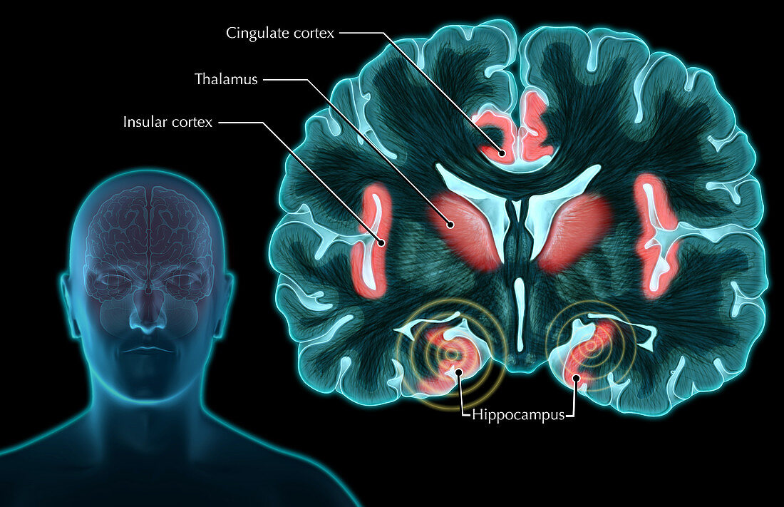
| An illustrated coronal (frontal) section of the brain,depicting areas of the brain which respond to pain,particularly in men. Typical regions of the brain,highlighted in red,which have demonstrated a response to painful stimuli are the thalamus (centre),insular cortex (outermost),cingulate cortex (top) and hippocampus (bottom). Recent studies have shown greater activity in the right and left hippocampus in men responding to pain,compared to women. The hippocampus has been known to respond to severe long-term traumatic stress | |
| Licence : | Droits gérés |
| Crédit: | Science Photo Library / Oto, Evan |
| Taille de l’image : | 5102 px × 3300 px |
| Model Release : | Non requis |
| Property Release : | Non requis |
| Restrictions : |
|
Prix pour cette image À partir de 45 €
Produit vendu
(Calendrier, Carte postale, Carte de vœux, Impression sur textile, Packaging etc)
À partir de 45 €
Usage commercial
(Affichage, Annonce presse, Annonce TV, Carte, Digital - hors rés. sociaux, Digital - rés. sociaux etc)
À partir de 45 €
Éditorial
(Digital, Journal, Livre, Livre pratique, Magazine, Télévision etc)
À partir de 60 €
Usage non-commercial
(Digital - hors rés. sociaux, Digital - rés. sociaux etc)
À partir de 120 €