Nerve Supply to the Disc
Numéro d’image : 12031753
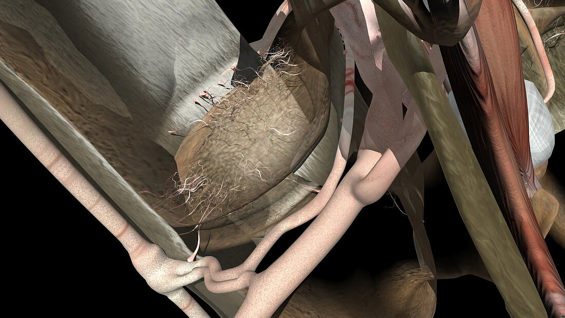
| This image highlights the extensive network of nerves that supply the perimeter of the lumbar disc. In the central portion of this image the lumbar disc has been removed. Remnants of small 'hair-like' projections are noted in the outer portion of where the disc was previously located. The top part of the bone endplate is the circular structure below these smaller nerves. The large anterior longitudinal ligament is noted to the left of the image and consists of parallel collagen fibres. In the foreground the spinal nerve root is seen to the right exiting the neuroforamen and dividing into the dorsal rami and ventral rami. Small muscles can be seen to the far right of the image | |
| Licence : | Droits gérés |
| Crédit: | Science Photo Library / Orthoclick |
| Taille de l’image : | 4552 px × 2561 px |
| Model Release : | Non requis |
| Property Release : | Non requis |
| Restrictions : |
|
Prix pour cette image À partir de 45 €
Produit vendu
(Calendrier, Carte postale, Carte de vœux, Impression sur textile, Packaging etc)
À partir de 45 €
Usage commercial
(Affichage, Annonce presse, Annonce TV, Carte, Digital - hors rés. sociaux, Digital - rés. sociaux etc)
À partir de 45 €
Éditorial
(Digital, Journal, Livre, Livre pratique, Magazine, Télévision etc)
À partir de 60 €
Usage non-commercial
(Digital - hors rés. sociaux, Digital - rés. sociaux etc)
À partir de 120 €
Mots clés
- anatomie,
- anatomique,
- art,
- colonne vertébrale,
- créé digitalement,
- dessin,
- humain,
- illustration,
- image créée par ordinateur,
- image de synthèse,
- ligament longitudinal antérieur,
- lombaire,
- médical,
- médicale,
- muscle,
- nerf,
- oeuvre,
- os,
- produit digitalement,
- racine du nerf,
- racine nerveuse,
- racines nerveuses,
- réalisé digitalement,
- système nerveux,
- système squelettique,
- vertébral