Muscle Fiber,TEM
Numéro d’image : 12019742
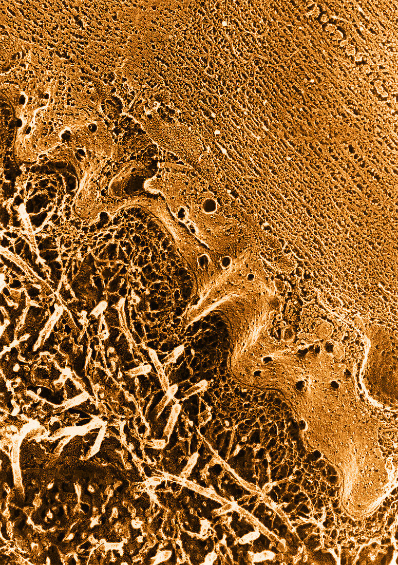
| Color enhanced transmission electron micrograph of a quick-frozen,deeply-etched preparation of skeletal muscle fiber and extracellular components. The upper right portion of the image is dominated by protein myofilaments. The basal lamina are the fine mesh structures on the outer edge of the cell membrane. The larger fibrils are collagen | |
| Licence : | Droits gérés |
| Crédit: | Science Photo Library / Fawcett, Don W. |
| Taille de l’image : | 3541 px × 5023 px |
| Model Release : | Non requis |
| Restrictions : |
|
Prix pour cette image À partir de 45 €
Produit vendu
(Calendrier, Carte postale, Carte de vœux, Impression sur textile, Packaging etc)
À partir de 45 €
Usage commercial
(Affichage, Annonce presse, Annonce TV, Carte, Digital - hors rés. sociaux, Digital - rés. sociaux etc)
À partir de 45 €
Éditorial
(Digital, Journal, Livre, Livre pratique, Magazine, Télévision etc)
À partir de 60 €
Usage non-commercial
(Digital - hors rés. sociaux, Digital - rés. sociaux etc)
À partir de 120 €
Mots clés
- basal lamina,
- cellulaire,
- cellule,
- collagène,
- couche limitative,
- couche limite,
- cryo-fixation,
- cryofixation,
- cryofixé,
- fibrillaire,
- fibrille,
- histologie,
- M.E.T.,
- membrane,
- membrane basale,
- MET,
- micrographie,
- micrographie électronique à transmission,
- microscope,
- microscope électronique à transmission,
- microscopie,
- muscle squelettique,
- myofilament,
- sarcolemme,
- science