Alzheimer's Disease,MRI
Numéro d’image : 12013642
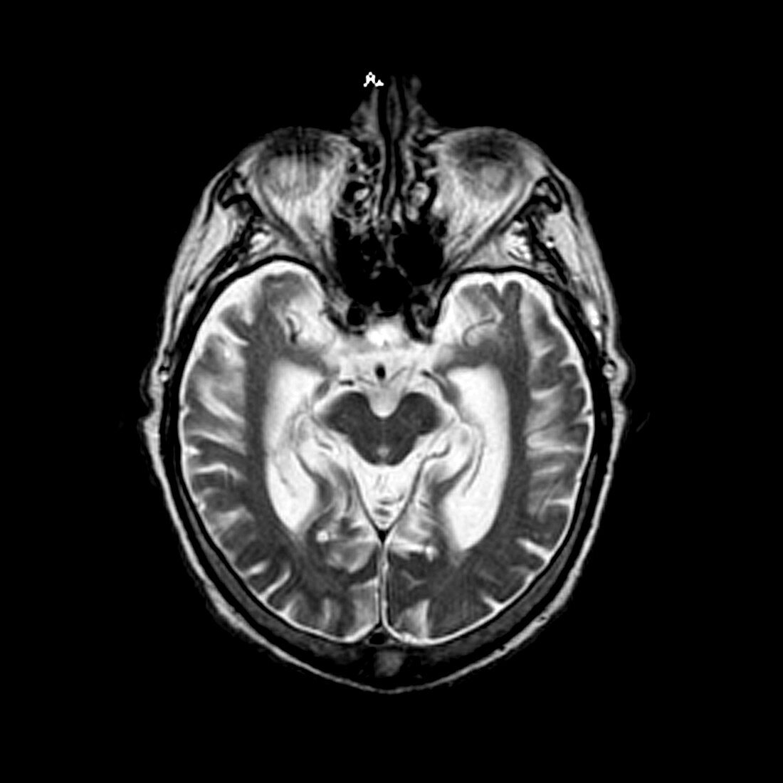
| This axial,(cross sectional),T2 weighted MRI image of the brain demonstrates severe atrophy of the medial temporal lobes,(hippocampal formations),with associated enlargement of the hippocampal/choroidal fissures due to the volume loss. This is a typical appearance seen with Alzheimer's disease | |
| Licence : | Droits gérés |
| Crédit: | Science Photo Library / Living Art Enterprises |
| Taille de l’image : | 3600 px × 3600 px |
| Model Release : | Non requis |
| Property Release : | Non requis |
| Restrictions : |
|
Prix pour cette image À partir de 45 €
Produit vendu
(Calendrier, Carte postale, Carte de vœux, Impression sur textile, Packaging etc)
À partir de 45 €
Usage commercial
(Affichage, Annonce presse, Annonce TV, Carte, Digital - hors rés. sociaux, Digital - rés. sociaux etc)
À partir de 45 €
Éditorial
(Digital, Journal, Livre, Livre pratique, Magazine, Télévision etc)
À partir de 60 €
Usage non-commercial
(Digital - hors rés. sociaux, Digital - rés. sociaux etc)
À partir de 120 €
Mots clés
- athrophique,
- atrophie,
- cérébral,
- cerveau,
- condition médicale,
- crâne,
- crânien,
- cranium,
- dégénératif,
- dégénérative,
- démence,
- dementia,
- désordre,
- état,
- I.R.M.,
- image de résonance magnétique,
- imagerie par résonance magnétique,
- imagerie par résonnance magnétique,
- IRM,
- lobe temporal,
- lobe temporal médian,
- maladie,
- maladie d'Alzheimer,
- médical,
- médicale,
- pathologie,
- système nerveux,
- système nerveux central,
- trouble