Heterotopic Gray Matter,MRI
Numéro d’image : 12010632
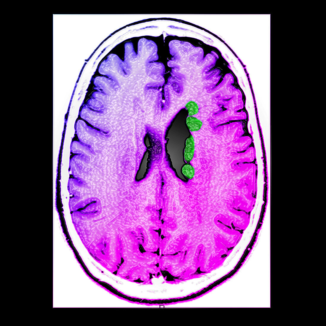
| This color enhanced axial (cross sectional) T1 weighted MRI image of the brain demonstrates nodules of heterotopic gray matter (green) along the margins of the left (on your right) lateral ventricle. This is a type of neuronal migration disorder. Cells destined to be in cortical gray matter begin in the subependymal regions in the germinal matrix zones. These neurons then migrate peripherally along radial-glial fibers to the cortex. If there is an insult during this process these neurons fail to migrate and will remain in the subependymal zones | |
| Licence : | Droits gérés |
| Crédit: | Science Photo Library / Living Art Enterprises |
| Taille de l’image : | 3600 px × 3600 px |
| Model Release : | Non requis |
| Property Release : | Non requis |
| Restrictions : |
|
Prix pour cette image À partir de 45 €
Produit vendu
(Calendrier, Carte postale, Carte de vœux, Impression sur textile, Packaging etc)
À partir de 45 €
Usage commercial
(Affichage, Annonce presse, Annonce TV, Carte, Digital - hors rés. sociaux, Digital - rés. sociaux etc)
À partir de 45 €
Éditorial
(Digital, Journal, Livre, Livre pratique, Magazine, Télévision etc)
À partir de 60 €
Usage non-commercial
(Digital - hors rés. sociaux, Digital - rés. sociaux etc)
À partir de 120 €
Mots clés
- amélioré,
- augmenté,
- cerveau,
- couleur,
- crâne,
- crânien,
- cranium,
- désordre neurologique,
- état,
- HETEROTOPIA,
- I.R.M.,
- image de résonance magnétique,
- imagerie par résonance magnétique,
- imagerie par résonnance magnétique,
- IRM,
- maladie,
- matière grise,
- médical,
- médicale,
- neurologique,
- pathologie,
- renforcé,
- tête