Heterotopic Gray Matter,MRI
Numéro d’image : 12010631
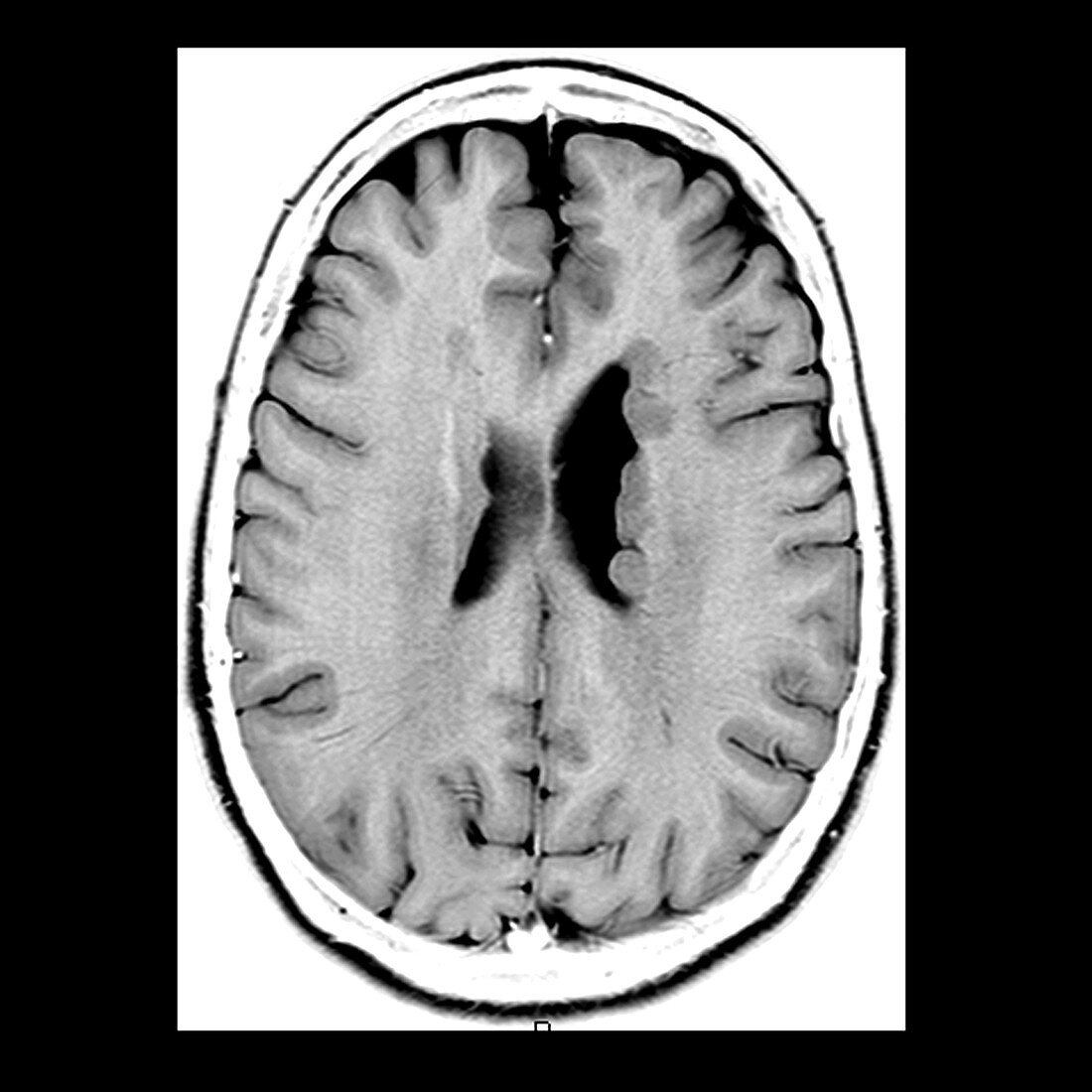
| This axial (cross sectional) T1 weighted MRI image of the brain demonstrates nodules of heterotopic gray matter along the margins of the left (on your right) lateral ventricle. This is a type of neuronal migration disorder. Cells destined to be in cortical gray matter begin in the subependymal regions in the germinal matrix zones. These neurons then migrate peripherally along radial-glial fibers to the cortex. If there is an insult during this process these neurons fail to migrate and will remain in the subependymal zones | |
| Licence : | Droits gérés |
| Crédit: | Science Photo Library / Living Art Enterprises |
| Taille de l’image : | 3600 px × 3600 px |
| Model Release : | Non requis |
| Property Release : | Non requis |
| Restrictions : |
|
Prix pour cette image À partir de 45 €
Produit vendu
(Calendrier, Carte postale, Carte de vœux, Impression sur textile, Packaging etc)
À partir de 45 €
Usage commercial
(Affichage, Annonce presse, Annonce TV, Carte, Digital - hors rés. sociaux, Digital - rés. sociaux etc)
À partir de 45 €
Éditorial
(Digital, Journal, Livre, Livre pratique, Magazine, Télévision etc)
À partir de 60 €
Usage non-commercial
(Digital - hors rés. sociaux, Digital - rés. sociaux etc)
À partir de 120 €
Mots clés
- cerveau,
- crâne,
- crânien,
- cranium,
- désordre neurologique,
- état,
- HETEROTOPIA,
- I.R.M.,
- image de résonance magnétique,
- imagerie par résonance magnétique,
- imagerie par résonnance magnétique,
- IRM,
- maladie,
- matière grise,
- médical,
- médicale,
- monochrome,
- N/B,
- NB,
- neurologique,
- noir blanc,
- noir et blanc,
- pathologie,
- tête