Brain,MRI
Numéro d’image : 12010612
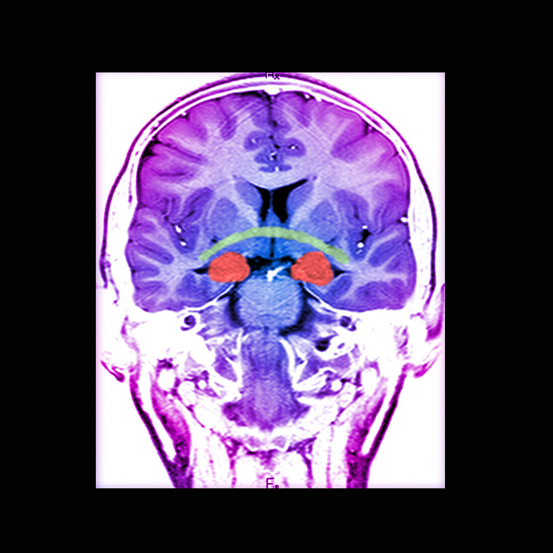
| This color enhanced coronal (frontal view) T1 weighted MRI image of the brain demonstrates normal cerebral anatomy. In this view you see the anterior commisure (green),which is a white matter tract connecting the temporal lobes. Also seen in this image is the amygdala (round red structures),located along the medial temporal lobes,just anterior and superior to the head of the hippocampus | |
| Licence : | Droits gérés |
| Crédit: | Science Photo Library / Living Art Enterprises |
| Taille de l’image : | 3600 px × 3600 px |
| Model Release : | Non requis |
| Property Release : | Non requis |
| Restrictions : |
|
Prix pour cette image À partir de 45 €
Produit vendu
(Calendrier, Carte postale, Carte de vœux, Impression sur textile, Packaging etc)
À partir de 45 €
Usage commercial
(Affichage, Annonce presse, Annonce TV, Carte, Digital - hors rés. sociaux, Digital - rés. sociaux etc)
À partir de 45 €
Éditorial
(Digital, Journal, Livre, Livre pratique, Magazine, Télévision etc)
À partir de 60 €
Usage non-commercial
(Digital - hors rés. sociaux, Digital - rés. sociaux etc)
À partir de 120 €