Lumbar Disc Herniation,MRI
Numéro d’image : 12010611
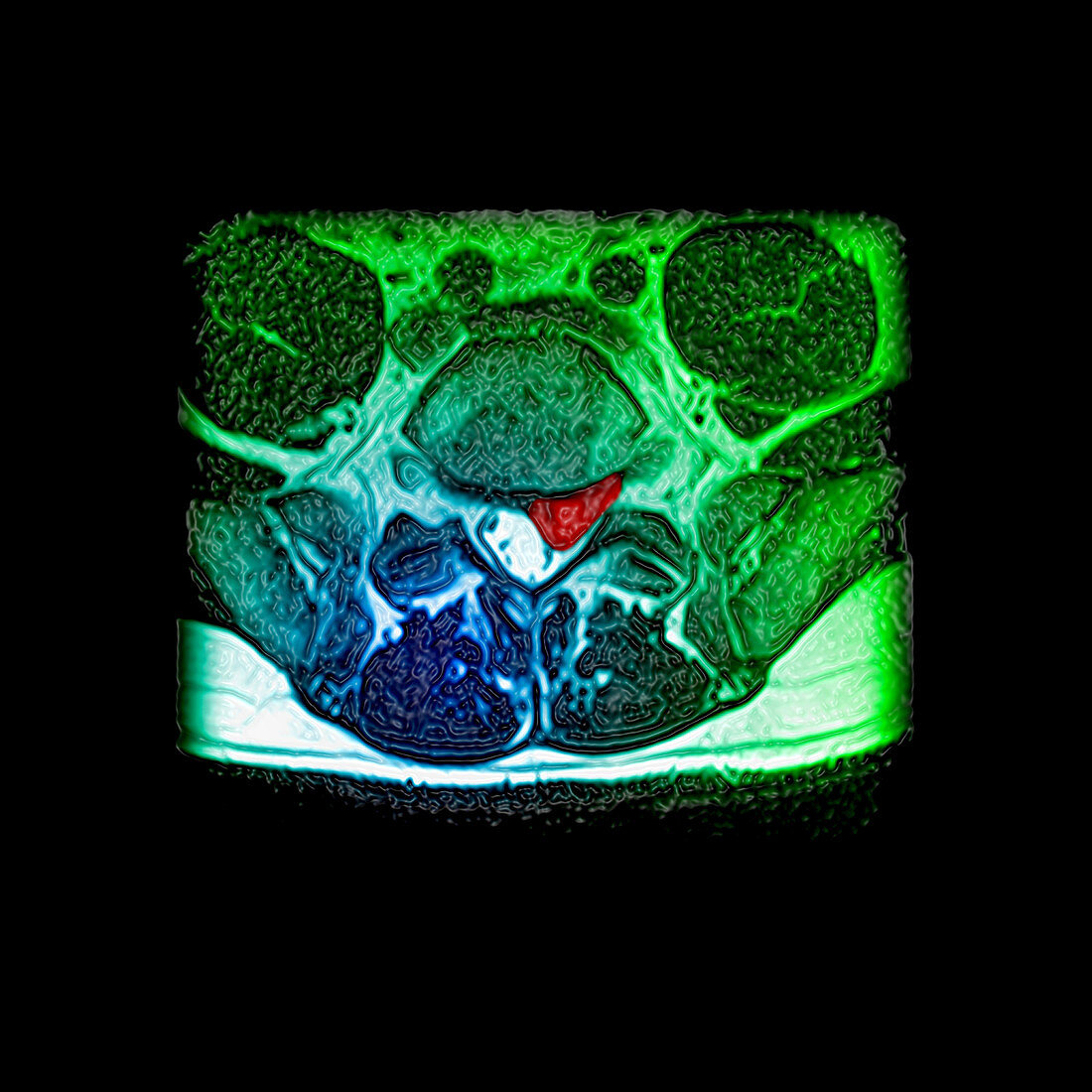
| This color enhanced axial (cross sectional) T2 weighted MRI image through the L5/S1 intervertebral disc level demonstrates a large left (on your right) sided herniation of the L5/S1 disc (red) compressing and displacing the S1 nerve root posteriorly | |
| Licence : | Droits gérés |
| Crédit: | Science Photo Library / Living Art Enterprises |
| Taille de l’image : | 3600 px × 3600 px |
| Model Release : | Non requis |
| Property Release : | Non requis |
| Restrictions : |
|
Prix pour cette image À partir de 45 €
Produit vendu
(Calendrier, Carte postale, Carte de vœux, Impression sur textile, Packaging etc)
À partir de 45 €
Usage commercial
(Affichage, Annonce presse, Annonce TV, Carte, Digital - hors rés. sociaux, Digital - rés. sociaux etc)
À partir de 45 €
Éditorial
(Digital, Journal, Livre, Livre pratique, Magazine, Télévision etc)
À partir de 60 €
Usage non-commercial
(Digital - hors rés. sociaux, Digital - rés. sociaux etc)
À partir de 120 €
Mots clés
- amélioré,
- arrière,
- augmenté,
- colonne vertébrale,
- couleur,
- de retour,
- disque,
- dos,
- état,
- herniation,
- hernie,
- hernié,
- hernie discale,
- I.R.M.,
- image de résonance magnétique,
- imagerie par résonance magnétique,
- imagerie par résonnance magnétique,
- IRM,
- L 5,
- L-5,
- L5,
- lombaire,
- maladie,
- médical,
- médicale,
- pathologie,
- rachis lombaire,
- renforcé,
- S1,
- vertebra,
- vertebrae,
- vertébral,
- vertèbre,
- vertèbres