Cavernous Malformation,MRI
Numéro d’image : 12010608
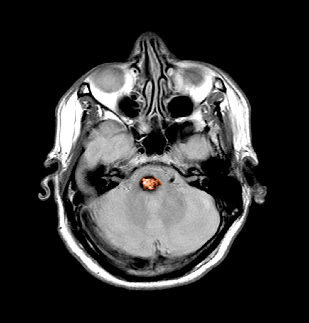
| This color enhanced axial (cross sectional) proton density weighted MRI image through the posterior fossa demonstrates the very typical appearance of a cavernous malformation within the pons (orange),which has also been called a cavernous angioma. This represents a slow flow type vascular malformation,which has also been referred to as an OCVM (occult vascular malformation) because they are generally angiographically invisible. These lesions represents dilated capillary spaces with no intervening normal brain. They can expand,however they are not usually associated with significant acute hemorrhages | |
| Licence : | Droits gérés |
| Crédit: | Science Photo Library / Living Art Enterprises |
| Taille de l’image : | 3600 px × 3762 px |
| Model Release : | Non requis |
| Property Release : | Non requis |
| Restrictions : |
|
Prix pour cette image À partir de 45 €
Produit vendu
(Calendrier, Carte postale, Carte de vœux, Impression sur textile, Packaging etc)
À partir de 45 €
Usage commercial
(Affichage, Annonce presse, Annonce TV, Carte, Digital - hors rés. sociaux, Digital - rés. sociaux etc)
À partir de 45 €
Éditorial
(Digital, Journal, Livre, Livre pratique, Magazine, Télévision etc)
À partir de 60 €
Usage non-commercial
(Digital - hors rés. sociaux, Digital - rés. sociaux etc)
À partir de 120 €
Mots clés
- amélioré,
- angiome,
- angiome caverneux,
- augmenté,
- cerveau,
- couleur,
- crâne,
- crânien,
- cranium,
- état,
- I.R.M.,
- image de résonance magnétique,
- imagerie par résonance magnétique,
- imagerie par résonnance magnétique,
- IRM,
- lésion,
- maladie,
- malformation caverneuse,
- médical,
- médicale,
- pathologie,
- Pons,
- renforcé,
- tête,
- tronc cérébral