Perineural Spread of Nasopharyngeal Carci
Numéro d’image : 12010575
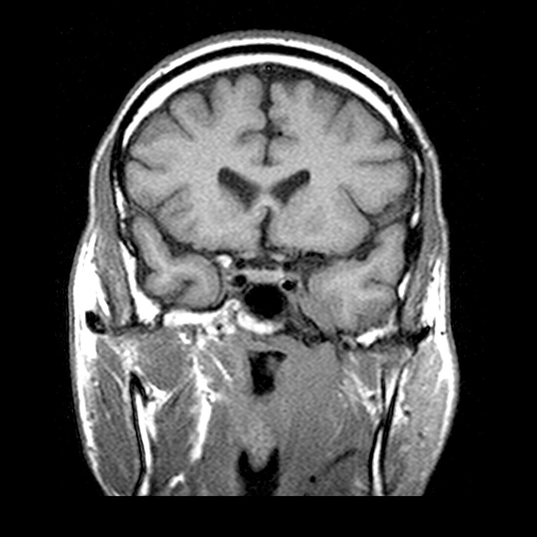
| A coronal (frontal) T1 weighted MRI (without contrast) through the nasopharynx and skull base. The scan shows a left sided (on your right) nasopharyngeal squamous cell carcinoma with extension into the masticator space and perineural spread along the third (mandibular) division of the trigeminal nerve | |
| Licence : | Droits gérés |
| Crédit: | Science Photo Library / Living Art Enterprises |
| Taille de l’image : | 3600 px × 3600 px |
| Model Release : | Non requis |
| Property Release : | Non requis |
| Restrictions : |
|
Prix pour cette image À partir de 45 €
Produit vendu
(Calendrier, Carte postale, Carte de vœux, Impression sur textile, Packaging etc)
À partir de 45 €
Usage commercial
(Affichage, Annonce presse, Annonce TV, Carte, Digital - hors rés. sociaux, Digital - rés. sociaux etc)
À partir de 45 €
Éditorial
(Digital, Journal, Livre, Livre pratique, Magazine, Télévision etc)
À partir de 60 €
Usage non-commercial
(Digital - hors rés. sociaux, Digital - rés. sociaux etc)
À partir de 120 €
Mots clés
- anatomie,
- anormal,
- cancer du cou,
- carcinome épidermoïde,
- cerveau,
- cou,
- I.R.M.,
- image de résonance magnétique,
- image médicale,
- imagerie médicale,
- imagerie par résonance magnétique,
- imagerie par résonnance magnétique,
- IRM,
- médecine,
- médical,
- médicale,
- nasopharynx,
- neurologie,
- neurologique,
- science,
- tête