MR of Malignant Brain Tumor,3 of 3
Numéro d’image : 12010570
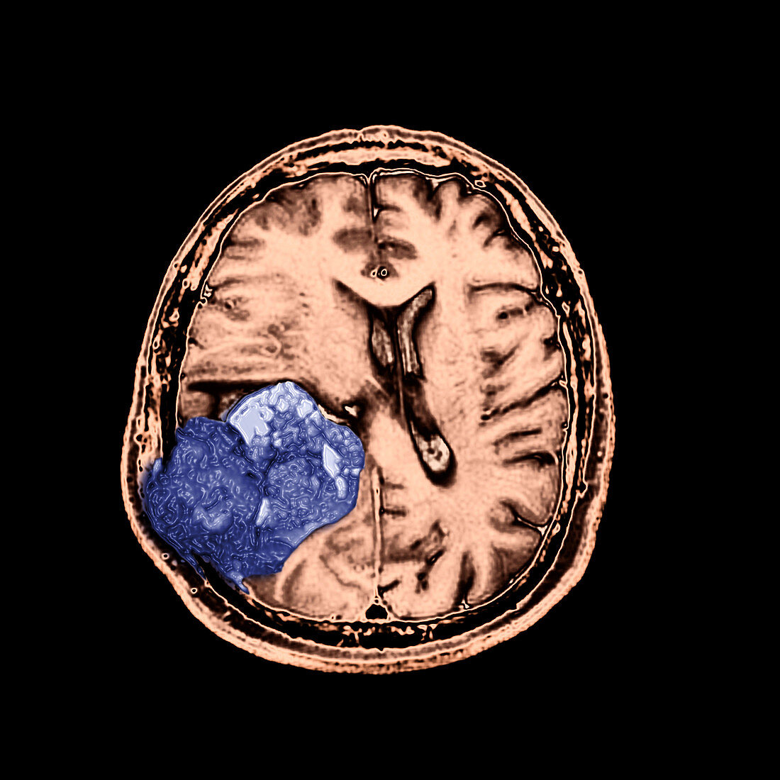
| This color enhanced axial (cross sectional) T2 weighted image of the brain demonstrates a heterogenous tumor (blue) in the right (your left) parietal lobe which destroys the overlying calvarium (skull bone) and extends into the overlying scalp. This tumor represents a malignant form of an astrocytoma. It is a WHO grade IV tumor called a Glioblasstoma Multiforme (GBM). These are most commonly seen in the elderly and unfortunately are one of the most common astrocytomas. They carry a poor,short term survival. Image 3 of 3 | |
| Licence : | Droits gérés |
| Crédit: | Science Photo Library / Living Art Enterprises |
| Taille de l’image : | 3600 px × 3600 px |
| Model Release : | Non requis |
| Property Release : | Non requis |
| Restrictions : |
|
Prix pour cette image À partir de 45 €
Produit vendu
(Calendrier, Carte postale, Carte de vœux, Impression sur textile, Packaging etc)
À partir de 45 €
Usage commercial
(Affichage, Annonce presse, Annonce TV, Carte, Digital - hors rés. sociaux, Digital - rés. sociaux etc)
À partir de 45 €
Éditorial
(Digital, Journal, Livre, Livre pratique, Magazine, Télévision etc)
À partir de 60 €
Usage non-commercial
(Digital - hors rés. sociaux, Digital - rés. sociaux etc)
À partir de 120 €
Mots clés
- anatomie,
- anormal,
- astrocytome,
- cancer du cerveau,
- cerveau,
- couleur améliorée,
- GBM,
- glioblastome,
- glioblastome multiforme,
- I.R.M.,
- image de résonance magnétique,
- image médicale,
- imagerie médicale,
- imagerie par résonance magnétique,
- imagerie par résonnance magnétique,
- IRM,
- lobe pariétal,
- malsain,
- médecine,
- médical,
- médicale,
- néoplasme du cerveau,
- neurologie,
- neurologique,
- science,
- tumeur cérébrale,
- tumeur maligne