'Leydig Cell Lysosomes,TEM'
Numéro d’image : 12007021
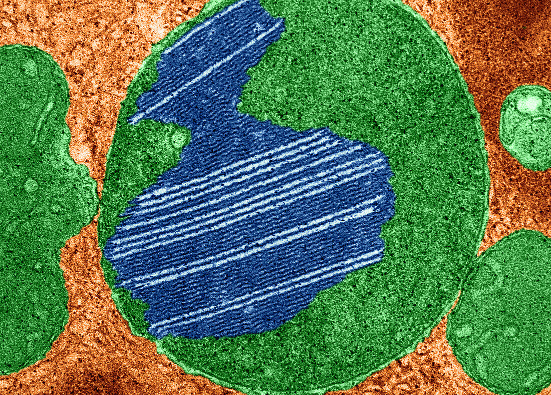
| Color enhanced transmission electron micrograph of lysosomes in the interstitial cells of Leydig in the testis of the domestic boar. Additional examples of laminated inclusions in lysosomes are illustrated here in micrographs from Leydig cells which,like other steroid-secreting cells,are rich in lysosomes. The clear spaces interposed among the lamellae are negative images of thin tabular crystals of unknown nature that have been dissolved in the course of specimen preparation. (Enhancement of 9C3323) | |
| Licence : | Droits gérés |
| Crédit: | Science Photo Library / Fawcett, Don W. |
| Taille de l’image : | 4302 px × 3087 px |
| Model Release : | Non requis |
| Property Release : | Non requis |
| Restrictions : |
|
Prix pour cette image À partir de 45 €
Produit vendu
(Calendrier, Carte postale, Carte de vœux, Impression sur textile, Packaging etc)
À partir de 45 €
Usage commercial
(Affichage, Annonce presse, Annonce TV, Carte, Digital - hors rés. sociaux, Digital - rés. sociaux etc)
À partir de 45 €
Éditorial
(Digital, Journal, Livre, Livre pratique, Magazine, Télévision etc)
À partir de 60 €
Usage non-commercial
(Digital - hors rés. sociaux, Digital - rés. sociaux etc)
À partir de 120 €
Mots clés
- cellulaire,
- cellule,
- cellule de Leydig,
- histologie,
- imagerie médicale,
- interstitiel,
- interstitielle,
- lamella,
- lamelle,
- Leydig,
- lysosome,
- M.E.T.,
- médical,
- médicale,
- MET,
- micrographie,
- micrographie électronique à transmission,
- microscope,
- microscope électronique à transmission,
- microscope électronique en transmission,
- microscopie,
- reproductif,
- sécréteur,
- sécrétion,
- sécrétoire,
- testicule,
- testicules,
- testis,
- testostérone