Chromatin Fibers from Chicken Erythrocyte
Numéro d’image : 12006994
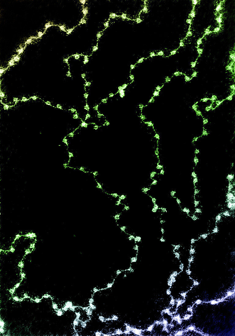
| Chromatin fibers spilling out of a chicken erythrocyte nucleus,lysed on a low ionic buffer. The fibrogranular appearance of chromatin in routine electron micrographs represents thin sections of a tangled mass of the fibers pictured here. Color Enhanced | |
| Licence : | Droits gérés |
| Crédit: | Science Photo Library / Fawcett, Don W. |
| Taille de l’image : | 2471 px × 3538 px |
| Model Release : | Non requis |
| Property Release : | Non requis |
| Restrictions : |
|
Prix pour cette image À partir de 45 €
Produit vendu
(Calendrier, Carte postale, Carte de vœux, Impression sur textile, Packaging etc)
À partir de 45 €
Usage commercial
(Affichage, Annonce presse, Annonce TV, Carte, Digital - hors rés. sociaux, Digital - rés. sociaux etc)
À partir de 45 €
Éditorial
(Digital, Journal, Livre, Livre pratique, Magazine, Télévision etc)
À partir de 60 €
Usage non-commercial
(Digital - hors rés. sociaux, Digital - rés. sociaux etc)
À partir de 120 €